Course Authors
Martin J. Carey, M.D., and L. Joseph Parker, M.D.
Release Date: 02/14/1999
Upon completion of this Cyberounds®, you should be able to:
Recognize the radiological findings from common conditions presenting to to an emergency department
Describe the anatomy of the bones of the hand and foot
List the components of the Glasgow Coma Scale.
A 24-year-old male with an inversion injury to the ankle presents with pain in the lateral aspect of the foot.
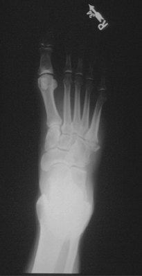
Q. What is the abnormality?
A. There is a fracture of the 5th metatarsal proximally. This is the commonest fracture of the lower limb but is often overlooked. The mechanism of injury is usually a sudden inversion injury as noted.
Q. What structure is inserted at this point?
A. The peroneus brevis tendon is inserted to the proximal end of the 5th metatarsal and avulses its origin.
Q. How can this injury be distinguished from a physeal plate in younger patients?
A. This fracture can be misdiagnosed in younger patients, where the physeal plate is still present. A fracture can be distinguished from the physeal plate, as the physeal plate is parallel to the long axis of the shaft of the 5th metatarsal, while a fracture is usually at 90° to the long axis. In older patients, accessory bones may also cause confusion. The accessory bones have rounded outlines, rather than the more ragged outlines seen in fractures. The classical accessory bones are called the os peroneum in the peroneus longus tendon and the os Vasalianum in peroneus brevis Note that, occasionally, there may be a fracture that involves both the metaphysis and the epiphysis. Separation of the epiphysis can also occur.
Q. What is a Jones fracture?
A. A Jones fracture is not associated with an inversion injury. It occurs in athletes, especially when they are increasing their training schedules. It has features suggestive of a stress fracture. The fracture occurs in the proximal aspect of the 5th metatarsal shaft. This fracture can be difficult to manage, as non-union is common.
Q. Name the bones of the foot!
A. The bones of the foot are: the calcaneus, talus, navicular, cuboid and the medial, middle and lateral cuneiforms, together with the metatarsals and the phalangeals.
Patient #2
This 18-year-old man states that he accidentally bumped his hand against the edge of a table.

Q. What abnormality is present?
A. There is a fracture of the 5th metacarpal. Approximately 20° of angulation is present. This degree of angulation should probably be accepted and either a simple dorsal slab applied, or else a 'boxing glove' cast.
Q. Do you think his story is credible?
A. This injury is classically associated with a closed fist striking a surface. A direct blow to the side of the hand is not likely to cause this injury.
Q. What other injury do you need to be careful to look for?
A. As the object struck is often someone's face, it is important to be sure there is no other associated injury to any of the metacarpo-phalangeal joints. Particularly, the presence of a laceration over one of the metacaro-phalangeal joints from a tooth needs to be carefully searched for. This injury, the 'closed fist laceration,' is very prone to infection and needs to be treated aggressively, with debridement and intravenous antibiotics.
Patient #3
This six-year-old child was brought in by her parents after they found her sitting on the floor playing with some coins. She had one coin in her mouth.

Q. What is the abnormality?
A. A coin is present in the distal esophagus. There are five areas where a foreign body is classically described as getting stuck in the esophagus. These are the cricopharyngeal narrowing at C6, the thoracic inlet at T1, the area where the aortic arch crosses the esophagus at T4, the bifurcation of the trachea at T6 and the distal esophagus at the hiatal narrowing at T10/11.
Q. What are the possible complications?
A. Possible complications of a foreign body in the esophagus include:
- Airway obstruction
- Stricture formation
- Perforation producing:
- Mediastinitis
- Cardiac tamponade
- Paraesophageal abscess
- Aortotracheoesophageal fistula
Q. How could this be treated?
A. The basis of management does not necessarily involve immediate removal of the coin but rather ensuring the passage of the coin into the stomach and through the pylorus. Once through the pylorus, the vast majority of all foreign bodies will pass through the GI tract safely. However, foreign bodies retained in the esophagus are a potential cause of the complications listed above. Possible management in this case, where the ingestion occurred within hours of presentation, may include:
- Repeating the chest film at two hours to assess spontaneous movement of the coin.
- The use of glucagon to relax the distal esophagus is well described in adults. This agent acts, specifically, on smooth muscle and, thus, is not useful for foreign bodies in the upper esophagus (where there is primarily striated muscle). Recently, there has been debate about the effectiveness of this agent in the disimpaction of foreign bodies and a controlled trial revealed no significant difference between the use of glucagon and placebo in the number of foreign bodies passed.(1) The use of glucagon in children is not well described.
- If the foreign body is known not to have been present for longer than 12-24 hours, then a Foley catheter may be passed distal to the obstruction, ideally under fluoroscopic control, the balloon inflated and then, gently, the catheter is removed. There is a small risk of aspiration using this technique and facilities should be immediately available for airway control. This technique should probably be a secondary option if direct endoscopic visualization and removal is not readily available. If available, then direct endoscopic visualization and removal is the ideal treatment for impacted foreign bodies in the esophagus which do not pass spontaneously after a short period of observation, or in those present over 12 to 24 hours.
Patient #4
This 25-year-old male has a history of night sweats, fever and increasing shortness of breath. He is homeless and lives on the streets. He denies drug abuse but you note that he has track marks on his arms. This is the first chest x-ray he has ever had.

Q. What is the differential diagnosis here?
A. The differential diagnosis in this case, with fibrosis and a cavitating lung lesion in the upper lobe on the right, should include:
- Tuberculosis
- Chronic necrotising pulmonary aspergillosis
- Cryptococcus
- Atypical mycobacterial infection
- Carcinoma
- Lung abscess
Q. What diagnostic tests would you arrange next?
A. A Mantoux test (0.1ml of purified protein derivative {PPD}) should be placed in a forearm. The test should be read at 48-72 hours after injection. Other investigations that may be considered acutely in the emergency department include the use of acid fast (Ziehl-Neelsen) staining of sputum or of a gastric aspirate. This patient, who is at high risk from HIV infection, should have this possibility explored. Culture of tissue, sputum or other body fluids usually takes some weeks to come back and so is not useful in the acute situation. The bottom line here is that diagnosis of TB in the emergency department can be difficult. It is important for the emergency physician to consider this diagnosis.
Let us say that this is not a 25-year-old homeless man but a 25-year-old 'Master of the Universe' who decided to blow some of his $1 million Christmas bonus from his stock trading on a holiday to South East Asia with his latest girlfriend. While in Asia, he prided himself on enjoying the local cuisine and was particularly fond of the shellfish. Since his return to the States, he has had pleuritic chest pain, fever and sweats and this morning had an episode of hemoptysis, hence his appearance in your emergency department.
Q. What is the possible diagnosis now and what is the organism responsible?
A. This patient is at risk of having acquired an infection from undercooked crayfish or freshwater crabs. Particularly in South East Asia, these shellfish harbor the metacercial form of the lung fluke (Paragonimus westermani). The larval worm hatches in the human stomach, migrates through the stomach wall, through the peritoneum, across the diaphragm and into the lung. The mature adult worms then produce local necrosis in the lung (and, occasionally, in the brain and liver, too). Cavitation and local necrosis occur and hemoptysis is not uncommon. Chest x-ray is often confused with tuberculosis. Eosinophilia is common. The diagnosis can be made from sputum and stool examination. Drug treatment is with praziquantel (Biltricide®) and bithionol.
Patient #5
This 25-year-old medical student presents with a two-day history of severe sore throat. On further questioning, it appears that he has experienced some weight loss recently (which he put down to his hard schedule) and also some night sweats over the past two weeks. He has a fever of 102°F and evidence of exudative tonsillitis. A zealous intern orders the chest X-ray.
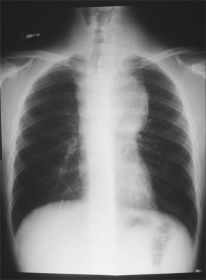
Q. What is the differential diagnosis in this case?
A. There is significant widening of the mediastinum. The differential diagnosis may be remembered using the mnemonic 'the fivet's':
- Thyroid (retrosternal goiter)
- Thymoma
- Teratoma
- 'Terrible' lymphoma
- Tortuous vessels
A more detailed list divides the mediastinum into three areas:(2)
- Superior Mediastinum
- Thymoma
- Retrosternal thyroid
- Zenker's diverticulum
- Anterior/Middle Mediastinum
- Dermoid cyst/teratoma
- Bronchogenic/pericardial cyst
- Morgagni diaphragmatic hernia
- Sarcoidosis
- Posterior
- Neurogenic tumors
- Bochdalek diaphragmatic hernia
- Achalasia, hiatus hernia
- Any mediastinal compartment
- Aortic aneurysm
- Lymphoma, teratoma
- Metastatic carcinoma
This young man had a lymphoma.
Patient #6
This 22-year-old female was involved in a road crash six days earlier. She described a three-minute loss of consciousness and suffered facial injuries from striking the dashboard. Her head CT scan was reported as normal and she had no facial fractures visible. In addition, she has her right leg in a cast from a fracture of the ankle. The fracture was managed with a closed manipulation under anesthesia and her facial lacerations were repaired. She was discharged on the second post injury day. She presents to the emergency department with a cough and a low-grade fever. The ER orders a chest X-ray.

Q. What is the abnormality apparent on the chest X-ray?
A. There is evidence of consolidation and some collapse of the right lower lobe. The cause of this problem is apparent on careful inspection. Have another look now if you do not see it.
A. This young lady with facial trauma has aspirated a tooth! It is visible as the white structure overlying the 8th rib next to the vertebral column. Aspiration of foreign bodies, often teeth, should be considered in anyone who has suffered facial injury, especially if associated with a loss of consciousness. Do not forget about dentures, either!
Q. How is this problem best managed?
A. This patient needs referral for a bronchoscopy for removal of the foreign body.
Patient #7
This 45-year-old man fell onto his outstretched hand from a height. He is complaining of pain in the wrist, with limitation of movement.

Q. What is the diagnosis?
A. This patient has a lunate dislocation. This is the commonest of the carpal bone dislocations. It is reported that the injury is missed about 40-50% of the time. Note that in a normal lateral view of the wrist, the 'cup' of the distal end of radius articulates with the proximal curve of the lunate, the distal curve of the lunate articulates with the capitate and the capitate articulates with the metacarpal. Each of these curved surfaces is in alignment. This alignment should always be checked for. In this example, the distal end of the radius does not articulate with the lunate. The lunate can be clearly seen, with the obvious 'crescent moon' shape that gives it its name, displaced to the palmar surface of the wrist. Note that on the anteroposterior view of the wrist (not shown), the lunate will resemble a 'piece of pie' (i.e., be a more triangular shape) in the case of a dislocation, whereas, normally, it will be closer in shape to a lopsided square.
Other dislocations of the carpal bones do occur. Injuries, where one or more of the proximal row of carpal bones remains in alignment with the radius, are identified using the prefix 'peri'. Examples of this type of injury include the perilunate dislocation -- the lunate stays in its normal alignment with the radius but all of the other carpal bones dislocate around it. Sometimes, the scaphoid may also remain (periscapholunate dislocation). Occasionally, one or more of the proximal row of carpal bones may fracture - a trans-scaphoid perilunate dislocation, for example. Other isolated carpal bones may dislocate. After the lunate, the scaphoid is the most commonly dislocated carpal bone.
Q. What is the treatment?
A. For patients suffering a dislocated lunate, closed reduction, under a general anaesthetic, is often successful. The wrist is held in supination and extension and traction applied. The operator palpates the lunate and uses their thumbs to push it posteriorly and distally. Once reduced, the wrist is returned to flexion and a short arm cast applied in slight wrist flexion. Failure of closed reduction is an indication for open reduction.
Q. What are four possible complications of this injury?
A. Complications of a lunate dislocation include:
- Late diagnosis, as noted above.
- Median nerve palsy. Rapid reduction of the dislocation usually resolves this problem, which is classically a neuropraxia. Again, delayed diagnosis makes this complication more serious.
- Sudeck's atrophy. This form of extreme osteoporosis, of unknown etiology, may occur after prolonged wrist immobilization. It is usually identified on cast removal, when the hand and wrist are warm, red, tender and painful. A radiograph reveals severe osteoporotic changes. Treatment of this complication includes analgesia and physiotherapy in the first instance.
- Avascular necrosis of the lunate is a potentially serious complication. All patients who have suffered a dislocated lunate should have regular radiographs (some authorities suggest monthly examinations for a period of six months after the injury) to identify early signs of this complication.
Q. What are the names of the bones of the hand?
A. The bones of the hand (Note: Bones are named starting laterally and proximally, and moving clockwise through the carpi) are:
In the proximal row: scaphoid, lunate and triquetral;
In the distal row: Hamate, capitate, trapezoid and the trapezium.
The pisiform bone overlies the triquetral.
Patient #8
This 28-year-old woman slipped on the ice. She is complaining of a very painful ankle with limitation of movement.

Q. What is the diagnosis?
A. This young lady has a fracture of the tibia at the ankle but also has a fracture of the fibula proximally. The take-home message here is that the whole of the fibula should always be examined whenever there is a history of injury around the ankle. This patient had so much pain in the immediate vicinity of the ankle fracture that she was unaware of the fibula injury below the knee until it was palpated.
Q. Whose name is associated with this type of fracture?
A. This injury is called a 'Maisonneuve fracture'.
Patient #9
The patient is a 42-year-old man assaulted during a fight at a bar. He was apparently struck on the head with a baseball bat. Although he had initial loss of consciousness, he is currently drowsy, but rousable, and complains of a severe headache. He smells strongly of alcohol. This is a CT of the head, without contrast, viewed with 'brain' windows.
Q. What abnormality is identified on these scans?
A. Left epidural hematoma
Q. How would this patient be managed?
A. Immediate neurosurgical intervention is required. Stabilize the patient per ATLS protocol, consider mannitol and hyperventilation with CO2 goal range of 35.

Q. What are the components of the Glasgow Coma Scale?
A. Glasgow Coma Scale
| Infant | Child/Adult |
| Eye Opening | |
| 4 Spontaneously | Spontaneously 4 |
| 3 To speech | To speech 3 |
| 2 To pain | To pain 2 |
| 1 No response | No response 1 |
| Best Verbal Response | |
| 5 Coos, babbles | Oriented 5 |
| 4 Irritable, cries | Confused 4 |
| 3 Cries to pain | Inappropriate words 3 |
| 2 Moans, grunts | Incomprehensible 2 |
| 1 No response | No response 1 |
| Best Motor Response | |
| 6 Spontaneous | Obeys commands 6 |
| 5 Localizes pain | Localizes pain 5 |
| 4 Withdraws from pain | Withdraws from pain 4 |
| Decorticate posture | Decorticate posture |
| 3 (flexion) | (flexion) 3 |
| Decerebrate posture | Decerebrate posture |
| 2 (extension) | (extension) 2 |
| 1 No response | No response 1 |
| TOTAL | TOTAL |
If GCS < 8? Intubate!
Patient #10
This 5-year-old child presents with a two-day history of a high fever and complaints of a sore throat. On the morning of admission, she had been noted to be increasingly uncomfortable and was refusing all food by mouth. On arrival, the child is sitting up and looks to be unwell. She will not talk to you and seems reluctant to swallow. Her temperature is 104°F. With the child sitting on the mother's lap, a soft tissue lateral X-ray is performed in the resuscitation room.

Q. What is the diagnosis here?
A. This child has a very large retro-pharyngeal abscess. This is evidenced by the significant swelling, visible anterior to the cervical spine. The epiglottis is normal.
Q. What age group seems most at risk from epiglottitis these days in the United States?
A. Epiglottitis used to be considered a disease of children. However, over the past few years, it appears that the incidence in children has declined markedly. This is probably due to the introduction of Haemophilus vaccination. In 1993, the ratio of cases of epiglottitis was 0.4 children to each adult. This compares with a ratio of 2.6 children to each adult in 1980. Interestingly, the mortality rate for adults remains at about 6-7%, while that for children has fallen to less than 1%. Classically, the adults are aged 42-47 and the male to female ratio is reported to be between 1.8 to 1 and 4 to 1.
Q. What organisms might you expect to identify in this patient?
A. Polymicrobial streptococcus species, anaerobes, Eikenella corrodens.
Q. What treatment would you initiate?
A. Penicillin G, 24 million units, continuous infusion or divide Q4-Q6h IV and metronidazole (Flagyl®), 1.0 gm load, and then, 0.5 gm Q6H IV (or cefoxitin 2.0 gm Q8H IV).