Course Authors
Lynn Schrader, M.D., and Martin J. Carey, M.D.
Dr. Lynn Schrader graduated from the University of Tennessee School of Medicine in 1987 and completed residency in emergency medicine at the University of Arkansas in 1990. She is board certified in emergency medicine and is currently an Assistant Professor of Emergency Medicine at the University of Arkansas where her focus is on EMS, disaster medicine and trauma care.
Drs. Schrader and Carey report no commercial conflict of interest.
Estimated course time: 1 hour(s).

Albert Einstein College of Medicine – Montefiore Medical Center designates this enduring material activity for a maximum of 1.0 AMA PRA Category 1 Credit(s)™. Physicians should claim only the credit commensurate with the extent of their participation in the activity.
In support of improving patient care, this activity has been planned and implemented by Albert Einstein College of Medicine-Montefiore Medical Center and InterMDnet. Albert Einstein College of Medicine – Montefiore Medical Center is jointly accredited by the Accreditation Council for Continuing Medical Education (ACCME), the Accreditation Council for Pharmacy Education (ACPE), and the American Nurses Credentialing Center (ANCC), to provide continuing education for the healthcare team.
Upon completion of this Cyberounds®, you should be able to:
Discuss the pathophysiology of rhabdomyolysis and of renal failure secondary to rhabdomyolysis
Describe the physical findings and laboratory abnormalities in rhabdomyolysis
List the treatment for patients with rhabdomyolysis
Discuss the unusual occurrence of upper extremity deep vein thrombosis (DVT) and its relationship to Thoracic Outlet Syndrome (TOS).
First Visit
It is a busy Monday afternoon in the ED and the next chart waiting is that of a 28-year-old female who complains of bilateral upper extremity pain and swelling over the last three days. She states that she did over 170 pushups as a part of a physical training program the day before the onset of her symptoms and has taken Tylenol® without relief. Today, she states that she has also noted nausea without vomiting and decreased oral intake. She has only urinated twice in the last 24 hours and noted a brownish discoloration to her urine. She denies any other symptoms and states that she has been in good health up until this episode. She does not drink or smoke and takes no routine medicines.
Physical exam reveals a slender female appearing in no distress. Her vital signs are all within normal limits with no orthostatic changes. On extremity exam, you note that both upper arms and forearms are edematous and tender on palpation. There is no erythema or warmth noted. Pulses are 2+ and equal and the neurological exam is normal. There is full active range of motion at all joints. The remainder of the physical exam is normal.
The initial dip urine was positive for large blood and 2+ protein with a specific gravity of 1.030. Nitrite and leukocyte esterase were negative. Microscopy showed 0-2 RBC, otherwise negative.
Q. What condition do you suspect based on the information given above?
A. Total CPK
Q. How would you treat this condition?
A. Hydration with NS to maintain a urine output of at least 200 ml/hr.
Further laboratory evaluation showed a Na+ of 142, K+ of 4.2, Cl- of 105, HCO3- of 24, BUN of 8 and a Cr of .9. The blood count showed a WBC of 8.9, Hgb of 13.8, Hct of 41.7 and PLT count of 211. The CPK was elevated to 65,890.
What Are the Pathophysiological Mechanisms That Occur with Rhabdomyolysis?
Rhabdomyolysis occurs when there has been injury to muscles leading to excessive muscle breakdown. The pathophysiology of muscle injury is thought to involve elevation of intracellular sodium and calcium. Normal skeletal muscle contains proteases and other enzymes that function at low levels of activity to decompose myofibril proteins. The activity of these enzymes is increased as the concentration of intracellular calcium rises, causing increased myofibril breakdown and the resultant release of cellular contents including myoglobin into the bloodstream.
What Are Some of the Causes of Rhabdomyolysis?
There are many causes of skeletal muscle injury or excess muscle breakdown. Direct injury to muscles can accompany multiple trauma, burns, external compression, prolonged immobilization, and compartment syndrome. Excess muscle activity can deplete muscle cell membranes and lead to breakdown of tissue. This is commonly seen in athletes who have overextended themselves, such as our patient, but it can be seen in many medical conditions as well. Status epilepticus, status asthmaticus, delirium tremens, neuroleptic malignant syndrome, agitation from psychosis or stimulant use, and thyroid storm can significantly increase a patient's muscular activity. Prolonged immobilization can result in local tissue damage from external compression. This is a common cause of rhabdomyolysis in the elderly, alcoholics and drug users. Other conditions associated with rhabdomyolysis include diabetic ketoacidosis (DKA), infections, hyperthermia, hypothermia, and localized hypoxia, as can occur with vascular injury or occlusion.
How Does Rhabdomyolysis Cause Renal Failure?
Untreated, rhabdomyolysis often leads to acute renal failure secondary to acute tubular necrosis(ATN). Myoglobin itself is not thought to be a toxin. However, when large amounts of myoglobin are excreted in acidic urine, myoglobin dissociates into two components -- globin and ferrihemate. Ferrihemate is currently suspected to be the toxic component of myoglobin that results in renal injury. Tubular obstruction by myoglobin probably plays a secondary role in rhabdomyolysis induced renal failure. While we are not able to predict with certainty which patients with rhabdomyolysis will develop ATN, the amount of elevation in CPK, K+, and PO4- are thought to have some degree of positive predictive value.
What Are the Usual Signs and Symptoms of Rhabdomyolysis?
Presenting signs and symptoms of rhabdomyolysis are often subtle and may be obscured by the underlying illness or trauma. Patients may complain of generalized or localized muscle soreness, weakness or swelling, or of a brownish colored urine. Physical exam may reveal swollen, tender muscles.
What Methods Are Available to Diagnose Rhabdomyolysis?
The diagnosis is usually made based on laboratory studies. Urine myoglobin was once the definitive test for rhabdomyolysis -- the urine will dip positive for blood with a negative microscopy if myoglobin is present. However, myoglobin is cleared from the plasma within as little as three to six hours post injury, resulting in a false negative result. Total CPK is now the recommended test for rhabdomyolysis. CPK is elevated immediately with muscle injury. Elevation of five times the normal value is thought to herald rhabdomyolysis and increasing CPK levels are thought to correlate with more severe disease. Peak CPK levels occur around 36 hours post injury. Other laboratory abnormalities seen with rhabdomyolysis include hyperkalemia, hyperphosphatemia, hypocalcemia, elevation of the BUN and anion gap.
What Is the Treatment for Rhabdomyolysis, and Why?
The treatment for rhabdomyolysis, saline hydration, is aimed at preventing ATN and renal failure. The increased fluids promote tubular flow, allowing the kidneys to filter the myoglobin and its breakdown compounds safely. Alkalization of the urine with sodium bicarbonate is also recommended to decrease the breakdown of myoglobin into its toxic products in the urine. Urine output should be kept at 200-300 ml/hr. Electrolytes and renal function should be closely monitored. If urine output can't be maintained with fluids alone, mannitol and Lasix® (furosemide) can be used to attempt to increase urine output. Diuretics are not always successful and dialysis may be needed in cases of renal failure unresponsive to the above therapy. In most cases, these patients should be admitted and monitored until CPK levels have fallen and renal function assured.
Hospital Course
The patient was hospitalized on IVF of D5 1/2 NS with one amp NaHCO3 at a rate adjusted to maintain a urine output of >200ml per hr. Her BUN and Cr remained normal and her CPK fell throughout her hospitalization. She was discharged after two days to continue oral fluids at home.
Second Visit
The day following her discharge, the patient returned to the ED complaining of swelling and tingling in her left arm. She stated that she had done well the evening after discharge, but again noted swelling that morning in the left arm only. She denied any strenuous activity or any trauma to the left arm. She also denied any changes in urination, nausea, vomiting, chest or abdominal pain or fever.
Physical exam was significant for diffuse non-pitting edema and mild tenderness of the left upper extremity to the shoulder. Pulses were again 2+ and equal and the neurological exam of the extremity was normal. There was erythema and localized warmth noted. The remainder of the physical exam was unremarkable.
After review of the old chart, a CPK was ordered, which remained elevated at 22,463, This was, however, significantly lower than her last inpatient level of 38,392. Electrolytes and renal function studies were unchanged. D-dimer was positive at >1000. Her chest film is pictured below.
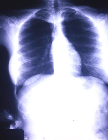
Q. What abnormality is seen on this film and what is its significance?
A. The abnormality is a left cervical rib.
Associated with Thoracic Outlet Syndrome
Q. What is your presumptive diagnosis?
A. Deep vein thrombosis (DVT)
Q. What test would you get to rule in or out that diagnosis?
A. Venous dopplers or venography
What Is "Effort Thrombosis?
Upper extremity DVT is rare, accounting for only about 1-2% of DVT. "Effort thrombosis" or Paget-Schroetter Syndrome occurs in young healthy adults after unusual or excessive use of the upper arm. It is commonly associated with Thoracic Outlet Syndrome (TOS). TOS may be due to anomalous fibrous bands in the costoclavicular area, cervical ribs or overdevelopment of the shoulder muscles. These conditions narrow the tunnel through which the brachial plexus, subclavian artery and subclavian vein pass causing pressure on these structures. This area can be further narrowed by shoulder attitudes, such as abduction, resulting in venous stasis and, ultimately venous thrombosis. Other etiologies of upper extremity DVT include catheter injury, tumors, clavicle fracture, CHF, and hypercoagulable states.
What Are the Physical Signs in Upper Extremity DVT?
Patients with upper extremity DVT may present with arm swelling, pain and cyanosis in the involved extremity. There may also be evidence of brachial plexus involvement if TOS is present. As with lower extremity DVT, diagnosis is made by venogram or duplex doppler study.
What Is the Treatment for an Upper Extremity DVT?
Standard treatment of upper extremity DVT is aimed at preventing propagation of the clot. Pulmonary emboli from upper extremity DVT rarely occur but should be considered in the management of these patients. Treatment is initially conservative -- rest, arm elevation and anticoagulation, beginning with IV heparin. Thrombolysis with streptokinase or urokinase has also been recommended. Many patients with effort thrombus as the etiology of their DVT will have continued symptoms after conservative treatment. Such patients should be referred to a surgeon for evaluation for resection of anomalous bands, cervical ribs or the first rib to prevent further thrombosis.
Outcome
Our patient underwent upper extremity doppler study which showed a thrombus extending from the brachial vein to the axillary vein. She was admitted and anticoagulated with heparin. Her swelling decreased and she was discharged on Coumadin (warfarin) to follow-up with vascular surgery.