Course Authors
Val Shestopalov, Ph.D.
Release Date: 08/28/2007
Upon completion of this Cyberounds®, you should be able to:
Describe the basic biology and physiology of the lens
Diagnose and classify cataracts
Conduct a systematic evaluation of patients with cataracts
Discuss the relationship between cataracts and systemic medical conditions.
The eye lens is a centerpiece of the refractive apparatus that provides accommodating power to focus vital visual information on the retina. This fascinating tissue is a living transparent matter with sophisticated morphology, which is achieved through the highly ordered development and the clearing away of light-scattering organelles from the bulk of lens fiber cells. The lens keeps growing throughout its entire lifespan and is designed in a unique way to preserve its transparency for decades. The transparent lens projects a sharp image on the retina. Alternatively, if the lens is cloudy from a cataract, the image will be blurred.
Cataract, or opacification of the lens, is the most common cause of visual impairment worldwide. Currently, no preventive therapy for cataracts exists and surgical removal is the only treatment option. Recent advances in our understanding of the genetics of human cataracts, in particular the inherited congenital form, together with the development of an array of animal models, have provided valuable new insights into normal vertebrate lens biology and the mechanisms that underlie cataract formation. The knowledge we now have about the mechanisms of hereditary cataract may also help us understand the manner in which environmental and nutritional factors act on the lens to promote opacification. All aspects of lens induction and development were comprehensively reviewed recently in the book Development of the Ocular Lens.(1)
In this Cyberounds®, we will briefly summarize the current knowledge of lens biology and discuss the relationship between some types of cataract and the underlying molecular causes.
Lens Biology and Physiology Relevant to Cataractogenesis
Cellular Organization and Tissue Transparency
The lens consists of two morphologically distinct cell types, an unremarkable cuboidal epithelium that covers the anterior surface and concentric layers of fiber cells that account for the remainder, and vast majority, of the tissue volume (Figure 1).

Figure 1. Structural Organization of Mammalian Lens.
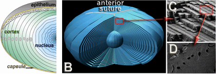
A. The bulk of the lens consists of concentric layers of fiber cells overlaid anteriorly by the epithelial sheath and encapsulated with thick basal membrane, the lens capsule. Progressive deposition of new fibers at the lens equator results in an onion-like architecture of the adult tissue shown schematically in B. Fiber cells retain full complement of their organelles only during elongation and maturation. After elongation is completed, these cells attach to the sutures and trigger ordered process of denucleation and organelle removal. Ultrastructural imaging shows details of fiber cell packing (C, top) and membrane interlocking (C, bottom), properties essential for tissue transparency and integrity during accommodation. (Courtesy of J. Kuszak)
The entire lens cell population is engulfed by a collagenous capsule, the thickest basal membrane in the mammalian body. The lens capsule is enriched in collagen IV, matrix proteins including laminin, fibronectin, heparin sulphate proteoglycan and entactin, which are laid throughout life by epithelial and fiber cells.(2) The capsule is thickest at the lens anterior and at the equator, the anchoring point of zonules.
The ciliary zonule consists of a series of fine fibers connecting the lens with ciliary muscle that stretches the tissue during accommodation. Mutation in the fibrillin gene, the major structural protein of these fibers, leads to the lens subluxation seen in Marfan syndrome. The lens epithelium forms an anterior subcapsular monolayer extending to the equator (Figure 1A). Epithelial cells (a population of about 500,000 per human lens) are typically described as cuboidal and retain proliferative capacity throughout life, are essential for the lens's continuous growth and the active epithelium function supporting the tissue homeostasis. These cells express alpha crystallins essential for their survival(3) but not the "lenticular" beta and gamma-crystallins; all epithelium is coupled by gap junctions formed by Cx43.(4)
The in vivo green fluorescent protein (GFP) tracing experiments showed that newly divided epithelial cells eventually migrate towards the equator and differentiate into fibers;(5) no cell turnover has been detected in the lens. Upon differentiation, epithelial cells start synthesizing a complement of lenticular proteins including beta- and gamma-crystallins, AQP0, Cx46 and Cx50 and elongate 1,000-fold to become fiber cells.
Lens fiber cells are uniquely shaped (Figure 1C), no more than a few micrometers wide but often exceeding a thousand micrometers in length.(6) In cross sectional profile they appear as flattened hexagons and their sharply angled membranes enclose a transparent cytoplasm that lacks the organelles found in regular cells.(7) Electron microscopy reveals elaborate interlocking cellular processes that help keep the entire mass of cells together in mechanically disturbed, fast accommodating lens tissue (Figure 1 C).
The tips of elongating fibers move towards sutures by growing over the surface of older fibers along posterior capsule and epithelial sheath at the anterior to form columns (Figure 1B). Fibers of the same growth shell have to travel different distances to reach sutures and, therefore, vary in length.(8) Fiber cell maturation is initiated after elongation is complete. This involves dramatic changes in the spectrum of expressed proteins. During maturation, fibers become compacted, their membranes tightly opposed and interlocked, leading to obliteration of the extracellular space. Along with increased crystalline content, these changes improve the refractive properties of the lens cortex and nucleus.
Fully differentiated, mature fibers in the lens center possess a combination of higher order membrane processes, as well as general cellular compaction, that weld fiber mass into a refractively distinct nucleus.(9),(10) The mature fibers are overlaid by layers of younger cells of the lens cortex. The outer layers of cortical fibers are nucleated and transcriptionally active, while deeper layers become devoid of nuclei and organelles.(11) As the cortex fibers mature, they progressively increase their refractive index, forming a gradient.(12) Slit lamp examination can distinguish up to 5 layers of optical discontinuity in the adult human lens cortex.(13)
Lens tissue transparency is programmed during development and cell differentiation and is achieved by combination of several factors: ordered cell organization, tight apposition of their membranes, macromolecular structure and homogeneity of cellular constituents. Highly regular arrangement and close apposition of lens secondary fibers result in minimal light scattering from destructive interference of cellular constituents at the wavelength of the visible spectrum.(14) Their cytozolic content becomes devoid of organelles and nuclei as they mature, leading to a high degree of homogeneity of the cellular constituents. Fiber cells synthesize and highly concentrate (up to 33% of dry weight) crystallins, proteins with chaperone activity that form high molecular aggregates of similar size, which remain soluble for decades.(15) Finally, lens homeostasis actively supports biochemical and structural stability over the lifetime. Disruption of either lens development or cellular mechanisms which support tissue homeostasis challenges transparency and causes opacification (i.e., a cataract).
Lens Development
The consecutive stages of differentiation direct transformation of regular cuboidal epithelium into these unusually shaped, perfectly ordered mature fibers with a remarkable reproducibility. Key cellular events, including induction of crystalline synthesis, rapid growth and elongation, followed by coordinated cleansing away of the organelles and membrane re-organization, are perfectly orchestrated to achieve the desired refractive properties of the "living glass." Unsurprisingly, alteration of either developmental stage inevitably compromises refined lens structure, challenges transparency and causes congenital or infantile type cataracts.(16),(17),(18)
The lens forms through temporally and spatially regulated patterns of induction and differentiation, coordinated by several transcription and growth factors. In the human, lens organogenesis (Figure 2) begins in the fourth week of gestation with thickening of the surface ectoderm overlying the developing optic vesicle to form the lens placode.(19) Invagination of this area produces the cup (lens pit), which closes over to form the lens vesicle that will pinch off from the ectoderm to become an independent tissue. A sub-population of epithelial cells lining the posterior wall of the vesicle rapidly elongate, obliterating the cavity of the vesicle to form primary lens fibers. Secondary lens fibers are subsequently produced throughout life by division of anterior lens epithelial cells in the equatorial zone of the lens and form concentric lamellae wrapping around more central fibers. All fiber cells are retained in the lens throughout life with minimal turnover of their protein constituents.
Figure 2. A Diagram Depicting Key Events of Lens Induction and Early Development.
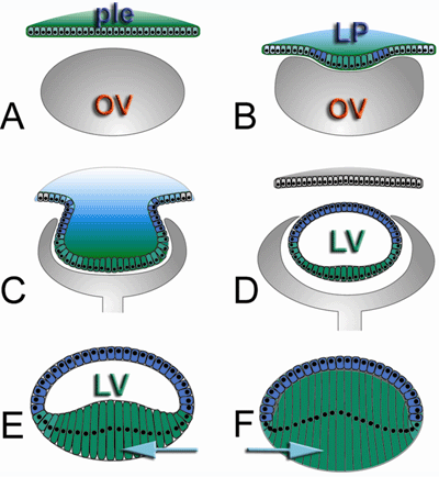
A. Contact between optic vesicle (OV) and presumptive lens epithelium (PLE) triggers thickening of the lens placode (LP, in B); C. Lens pit formation by invagination of the LP results in pinching off the lens vesicle (LV, in D,E). In the separated LV posterior cells (green) differentiate and elongate (E) to become primary fibers (F, arrows). Secondary fibers wrap around these cells continuously over the lifetime.
The development of lens structural properties is a function of cell proliferation, differentiation and de-nucleation. Quite uniquely, all three processes are continuously occurring in this tissue throughout life, which allows us to define four developmentally distinct zones.
Lens epithelium at the lens anterior pole retains proliferative capacity, thus comprising the germinative zone. Epithelial cells migrate towards the lens equator, withdraw from the cell cycle and elongate into fibers within the lens outer cortex, the elongative zone. When elongation is complete, fiber cells detach from the capsule and the epithelium, restructure their lateral membranes complexes forming the maturation zone within lens deep cortex. The final stage in the life of a fiber cell begins with the abrupt but organized loss of nuclei and intracellular organelles to produce the fully differentiated, matured fibers comprising the inner core of the lens termed nucleus.(7)
Major differentiation events occur in the germinative zone near the lens equator, where epithelium cells divide, undergo differentiation and move to a narrow transition zone, where they start elongation and become the fiber cells (Figure 3).
Figure 3. Epithelial Cells Differentiate into Fibers in the Germinative Zone at the Lens Equator and Start Elongation in the Transition Zone.
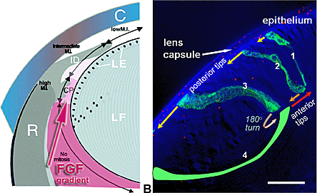
A. Growth factors (FGFs) gradient provide essential signaling the control the epithelial (LE) proliferation and differentiation. The highest mitotic index (MI) is detected at the lens equator (arrow). B. Youngest fibers were labeled with GFP protein to highlight cellular events at the beginning of elongation in the transition zone of the chicken lens. Growing fibers (1-2-3-4) move tips that will become posterior along the capsule (yellow arrows) and turn their to-be-anterior tips 180 degrees to move in the opposite direction along the epithelium-fiber interface (red arrows). Fibers 1, 2, 3 were labeled with GFP (green) delivered by gold particles bombardment (red dots).
Growth factors synthesized in the retina form a gradient near the lens equator, which has been shown to provide signaling essential for epithelial proliferation and differentiation.(20),(21),(22),(23) Fiber elongation occurs by coordinate movement of the opposing fiber cell tips along the posterior capsule and the epithelium-fiber interface (Figure 3B), forming layers of developmentally synchronized cells. Each layer will eventually form single "growth shell," wrapping the entire lens surface suture-to-suture.
Points at which tips of the secondary lens fibers come into apposition result in lines of optical discontinuity or 'sutures'. Sutures in the human lens resemble the letter "Y" in embryonic lenses but their architecture becomes progressively more complex by additional branching as lens grow and age.(8) The lens is surrounded by a capsule of mesenchymal origin and, during embryogenesis, blood is supplied from the hyaloid artery forming a temporary vascular bed, the tunica vasculosa lentis (TVL). The TVL is fully formed in 50 mm and regresses completely in the 160 mm human embryo but only postnatally in rodents.(19) Successful organogenesis results in a transparent biconvex lens suspended in the eye by zonular ligaments between the aqueous humour and the vitreous body.
Early lens development defects may be profound and cause total opacity or even lens dysgenesis. However, more benign forms are restricted to fetal or embryonic nuclei that become overlaid by normal fibers and have minimal effect on visual acuity.
A number of key transcription factors such as PAX6, PITX3, MAF, HSF, AP-2, SIX3, SIX5, SOX1, SOX2, CHX10, BMP7, and some others turn on early differentiation, controlling eye induction, formation and crystalline synthesis (Table 1). PITX3 (expressed in lens placode and forming lens pit),(24) AP-2 (lens vesicle),(25) SIX3 (expressed in the optic vesicle and anterior neural plate), SIX5 (expressed in the mature lens), BMP7 (lens placode)(26) and OPTX2 (expressed in lens placode and optic vesicle) also appear to play important sequential roles in the induction of eye and lens development.(27),(28)
Homozygous mutations in genes encoding these factors result in severe developmental abnormalities and are often lethal; heterozygous mutations show phenotypes varying from cataract to aphakia, aniridia, microphthalmia both in vertebrate animals and in humans (17),(29),(30) (Table 1). Signaling via growth factors, including IGF-1, multiple fibroblast growth factors (FGFs), BMPs, FOXE3 and LDGF, have been implicated in later stages of lens formation and fiber cell differentiation.(21),(31),(32) Functional inactivation of or mutation in these genes encoding these proteins results in severe lens cell dysgenesis, abnormal differentiation and is associated with various cataracts in animals and humans as shown in Table 1.(17),(33)
To conclude, the role of early regulators is crucial for normal lens development and alteration in these genes puts the lens structure and refractive properties at risk (Table 1). In turn, the presence of the developing lens appears to be crucial for the normal development and organization of other ocular tissues in the anterior chamber of the eye;(34) an aberrant or missing lens may cause dysgenesis of the whole eye.
Refractive Properties of the Lens
The continued peripheral growth of the lens, which results in the concentration of older tissue with higher crystalline content towards the center, has an important functional outcome -- producing a lens of variable refractive index.(35) The most important consequence of such organization implies that the biological lens is optically far superior to a glass bead of similar diameter. To illustrate this point, the refractive plane of a glass bead and a vertebrate (fish) lens were examined by laser polarimetry. As shown in Figure 4, when compared to a glass bead possessing significant variability in refractive plane (spherical aberration), the biological lens projected a nearly perfectly planar image, a property essential for adequate image projection on the retina and visual acuity.(36)
Figure 4. Laser Polarimetry of a Glass Bead and a Vertebrate Lens.

Laser polarimetry of a glass bead (A) and a vertebrate lens (B) of similar diameter demonstrating significant spherical aberration of light passing through the glass as compared to nearly perfect correction of the focal plane in biological lens. Yellow dots indicate focal points of images projected from different angles (Courtesy of J. Kuszak).
Lens cell physiology is entirely directed to the maintenance of the transparency-friendly architecture which serves to minimize photooxidative stress (via catalase, glutathione redox cycle and the mercaptopuric pathway), and changes in hydration and electrolyte imbalance. In contrast, metabolic activity is highest in the lens epithelium and young elongating fibers. These superficially located cells are metabolically coupled to the deeper metabolically inert layers via an elaborate system of gap junctions and cell-cell fusions, allowing water, ions and metabolite movement within the lens.(4),(37)
Fusion between neighboring cells occurs in deeper nucleated fibers and persists into the anucleated cortex and the nucleus, thus providing a putative pathway for macromolecular diffusion and limited protein turnover.(38) This arrangement represents a true syncytium and is thought to compensate for the lack of vascular supply to the lens cells.(39)
Lens Proteins, Physiology and Homeostasis
Lens refractive properties are dependent on the high concentration, solubility and multimer assembly of the major cytosolic proteins, crystallins. Crystallins are highly stable, water soluble proteins some of which are enzymes and potent molecular chaperones. In human lenses, three major families are represented by alpha-, beta- and gamma-crystallins.
While beta and gamma are closely related globular proteins characterized by four beta-sheets forming the "Greek key motif" (resembling the Greek ornament), alpha-crystallins are a distinct family of small heat shock proteins (sHSP, implicated in response to stress), which are essential for maintenance of lens protein solubility and prevent aggregation. Alpha-crystallin, a prominent member of sHSP family and a major structural protein of the eye lens, is a large polydisperse oligomer of two isoforms, alphaA- and alphaB-crystallins.
Numerous studies have demonstrated that alpha-crystallin functions like a molecular chaperone in preventing the aggregation of various proteins under a wide range of stress conditions.(40) The molecular chaperone function of alpha-crystallin is thus considered to be vital in the maintenance of lens transparency and in cataract prevention. Alpha-crystallin selectively interacts with non-native proteins, thereby preventing them from aggregation and helps maintain them in a folding competent state. It has been proposed and generally accepted that alpha-crystallin suppresses the aggregation of other proteins through the interaction between hydrophobic patches on its surface and exposed hydrophobic sites of partially unfolded substrate protein.(41),(42)
Members of the beta/gamma-crystallin superfamily comprise multiple proteins capable of forming compact globules with hydrophobic residues packed inside the Greek key motif-like (resembling the Greek ornament) central core.(43) Many mutations mapped to this crystallin superfamily were shown to be cataractogenic (Table 1). In particular, mutations in CRYAA lead to autosomal dominant and autosomal recessive congenital cataracts,(44) CRYBB2 gene harbors three such mutations(45),(46),(47) and CRYBA1 two mutations(48),(49) found in different ethnic backgrounds. CRYGD mutations affected solubility of gamma-crystallin gene, also causing cataracts.(50) Abnormal cleavage of gamma-crystallin in the lens nucleus has also been shown to be cataractogenic but was actually triggered by the absence of functional gap junction protein connexin.(50),(51),(52)
Since the alpha-crystallins belong to the family of sHSPs, it might be interesting to note that a mutation in a gene coding for heat shock transcription factor 4 (gene symbol HSF4) is associated with a dominant, lamellar cataract.(53) Crystallin genes CRYBB2 and CRYBA1 are the most affected by cataractogenic mutations. Homologous mutations in the mouse Crybb2 gene possess increasing severity of the phenotype, which is temporally correlated to the expression of the Crybb2 gene.(54) In the postnatal week, the characteristic bow configuration of the nuclei was altered, and swelling of the lens fibers occurred. Faint anterior opacities seen at postnatal day 15 are followed by sutural cataracts at day 25, nuclear cataract at 30 days, lamellar perinuclear opacities at 35 days, and total nuclear with anterior and posterior polar cataracts at 45 days.(55) Cataractogenesis is associated with an intralenticular increase in water, sodium and calcium, and a decrease in potassium, reduced glutathione, ATP and altered membrane permeability.
Among the lens cytoskeletal proteins, vimentin, filensin (CP115/CP95) and CP49 (phakinin), CP49 seems to be very important for the lens because it is the only one of the three proteins sensitive to the deletion or mutation of its gene. These proteins accumulate in mature fibers, while biosynthesis of vimentin and keratin ceases at this stage in the development. Beaded filament proteins were shown to associate with alpha-crystallins and facilitate the chaperone activity of alpha-crystallin assemblies. Mutations in CP49-encoding gene BFSP2 were shown to be responsible for dominant cataracts in humans;(56) their phenotypes, however, seem to be variable, ranging from congenital nuclear, sutural or stellate cataracts to juvenile-onset cataracts.
Lens Homeostasis Depends on Cell-Cell Communication
The lens of the eye is an avascular tissue, a solid cyst of cells, which grows throughout life by addition of new cells at the surface, while the older cells become buried by the newer generations. In contrast to the epithelial and young fiber cells that retain their cellular organelles and are exposed to the nutrients in the aqueous humors bathing the lens, these deeper lying cells rely on cooperation with the surface ones to maintain correct ion and metabolite concentrations within the cytoplasms such that the crystallins remain in solution and do not aggregate (cataract). To maintain this critical cooperation, the long-lived lens fibers are well interconnected via the two partially overlapping communication pathways: gap junctions, and recently discovered cell-cell fusions.(38),(57) While gap junctions represent classical transmembrane channels coupling all fiber and epithelial cells,(39) cell-cell fusions interconnect only the mature fibers in the lens core region.
The network of gap junctions joins the lens cells into a syncytium with respect to small molecules, permitting metabolic cooperation: intercellular diffusion of ions, metabolites and water. In some species, gap junctions account for more than 50% of the plasma membrane surface area.(58) Gap junctions are formed by a family of integral membrane channel-forming proteins called connexins. The unusual physiology and longevity of the lens fibers require the special set of connexins, Cx50 and Cx46, to couple together encapsulated cells.
Connexins are rather specific to the lens -- the Cx46 has not been detected in other tissues. A series of elegant experiments using transgenic "knockout" and "knockin" technologies showed that differentiation state-specific expression of these two connexins is essential for normal lens growth and transparency.(59),(60),(61) Mutations and functional inactivation of gap junction-mediated communication in the lens disconnect mature fibers in the lens center from metabolic support provided by peripheral cells and cause cataracts in human and animal lenses.(39),(51),(52),(62)
Recent experimental data obtained in vertebrate lenses suggest that a pathway that permits the cell-cell diffusion of macromolecules links lens fiber cells just before they degrade their organelles. The first evidence for the existence of this pathway has come from the ectopic expression of green fluorescent protein (GFP).(38),(57) Three-dimensional reconstruction of plasma membrane architecture of individual fibers revealed the presence of fusion pores between cells in the core of the chicken and mouse lenses. Such fusions represent conduits for the diffusion of cytoplasmic and membrane proteins in the lens core.
Fiber cell membrane fusions have been described in the lenses of several species(63),(64),(65) and appear to be particularly numerous once cells have reached the sutures. Assuming that fusions are a universal feature of the vertebrate lens, this raises the possibility that newly synthesized proteins could diffuse into the metabolically inert core of the lens. By allowing the intermingling of cytoplasmic components, fusions could equalize the refractive index in adjacent cells and thereby minimize internal light scattering within the tissue. Experimental inactivation of Lim2 protein, which is essential for cell-cell fusion in the mouse lens, caused nuclear cataracts and severe refractive abnormalities.(5) Significantly, mouse To3 mutation mapped to the Lim2 gene causes dense cataract,(66) while human mutation in LIM2 protein also leads to autosomal recessive presenile cataract formation.(67)
In addition to the proteins supporting cell-cell communication, lens membranes contain a complement of channel proteins supporting an exchange of ions and small molecules between the cytosol and the media. Lens membranes are particularly rich with water channels formed by aquaporin (AQP0/MIP26) protein, chloride channel proteins and recently characterized hemichannel pannexin1.(68),(69),(70) Mutations in AQP0 have long been known to cause cataracts in human and mouse lenses indicating the importance of water transport and, possibly, cell-cell adhesion functions assigned to this protein.(71),(72)
Summary
The development and growth of the lens throughout life rely upon the sequential time-limited expression of a number of genes. This highly ordered sequence guarantees the lens functional and structural stability, the foundation of life-long transparency, the major function of the tissue. Specialized soluble and membrane proteins expressed in the lens are essential to homeostasis of the lens, which is devoid of vasculature.