Course Authors
Richard W. Smalling, M.D., Ph.D.
Release Date: 11/30/1996
Background
Although isolated mitral stenosis is relatively rare in Western civilization, it still occurs and is associated with significant disability and morbidity. It remains more prevalent in Third World countries, particularly in Asia, Eastern Europe, Africa and the Middle East. Symptoms develop very slowly and typically patients present in their late 20's, although there is a wide variation and a bimodal distribution of presentation, with a late peak occurring in the 60's and 70's. Patients with isolated mitral stenosis, or mitral stenosis with only mild mitral regurgitation, who are New York Heart Association Class II - III would be considered candidates for mitral commissurotomy baring other significant valvular or coronary disease.
But, should the patient have a surgical commissurotomy or are there other less invasive approaches we need to consider? In order to answer our question, let's first briefly review the typical physical findings and laboratory evaluation.
Physical Findings
Patients with mitral stenosis are typically thin and female. They often have rales on chest examination which are non-positional. They have a discrete and non-displaced PMI with an opening snap heard in the tricuspid and mitral area and a low frequency diastolic rumble at the apex. If the mitral stenosis has been long-standing and accompanied by pulmonary hypertension, an increased P2 may be heard and a right ventricular lift may be appreciated along the left sternal border. Late in the course, patients begin to develop peripheral edema which may, as the right heart failure worsens, proceed to severe lower extremity edema and ascites.
Laboratory Evaluation
The chest x-ray usually demonstrates left atrial enlargement with pulmonary vascular redistribution and Kerly B-lines.
The ECG demonstrates right axis deviation with left atrial abnormality (so called P-mitrale) and, occasionally, right ventricular hypertrophy. Patients presenting relatively late in the course frequently are in atrial fibrillation.
Cardiac echo is essential for demonstrating left atrial size (usually large) and evaluating mitral leaflet mobility, calcification, sub-valvular apparatus and degree of thickening (see mitral valve scoring in Table 1). Additionally, doppler analysis can give accurate estimates of the pressure gradient across the valve, pulmonary artery pressure and degree of mitral regurgitation.
Historical Perspectives of Mitral Commissurotomy
Closed mitral commissurotomy was first described in 1923. The operative mortality associated with this procedure, as it has developed, ranges from 0-12% with an average of 4.8%. The 10-year survival after closed commissurotomy is 82%, with 66% of patients being event free at 10 years.
Open mitral commissurotomy was described in the early 1970's. The operative mortality for this procedure also ranges from 0-12% with an average of approximately 3.9%. The 10-year survival is 80-92% with 70% of patients being event free at 10 years.
Thus, both surgical procedures offer similar risks and outcomes. Now, let's return to our question, Is there any other therapeutic option for mitral stenosis that a physician can suggest to the patient?
Percutaneous Mitral Commissurotomy
In the mid-to-late 1980's percutaneous mitral valvuloplasty was developed. This technique required a transseptal technique involving puncture of the intra-atrial septum with a Brockenbrough needle and Mullins sheath. A balloon tipped catheter filled with hydrogen was then advanced through the Mullins sheath into the left atrium and left ventricle. Early on, this catheter was maneuvered to the apex and out the left ventricular outflow track and a relatively stiff wire was then placed through the transseptal catheter, left sided chambers and into the descending aorta. Subsequently, the technique was modified and wires were left in the left ventricular apex rather than in the descending aorta.
The intra-atrial septum was then dilated with an 8 mm angioplasty balloon and, in the majority of centers, two angioplasty balloons were placed across the mitral valve orifice via the trans-septal puncture. The balloons were inflated simultaneously with subsequent, commisural splitting. Additionally, hybrid balloons using multiple balloons on a single shaft were also developed and utilized in some European centers.
Which Patients Are Candidates for Mitral Valvuloplasty?
Valvuloplasty is sometimes overlooked as a viable treatment option even for patients who are reasonable candidates for this much less invasive procedure. Patients who have 2+ or less mitral regurgitation, New York Heart Association Class II - III symptoms of heart failure and mitral valve leaflets which are relatively pliable, minimally calcified and without significant subvalvular stenosis are candidates for mitral valvuloplasty.
Several groups have described scoring systems for the mitral valve. The most commonly utilized one in the United States is illustrated in Table 1, and was developed by investigators at the Massachusetts General Hospital. The mitral valve is evaluated using four categories, including mobility, subvalvular thickening, thickening of leaflets, and calcification. The grading system ranges from 1 to 4 in each category, giving a range of scores from 4 to 16.
Any patient with mitral stenosis who is without significant mitral regurgitation (less than 2+), and with echo scores less than or equal to eight, should be considered a candidate for mitral valvuloplasty. Patients with left ventricular end diastolic pressures less than 10 mm Hg are stronger candidates than those with higher left ventricular end diastolic pressures who must be considered at greater risk for adverse events. Similarly, patients who are New York Heart Association Class IV pose a greater risk for the procedure.

Table 1. Percutaneous Valvuloplasty Echo Scoring for Mitral Valve Stenosi.
| Characteristic | Description | Score |
|---|---|---|
| Mobility | Highly mobile valve, except tips; Mid and base move normally; Continued forward movements in diastole; Minimal forward leaflet movement | 1 2 3 4 |
| Subvalvular; Thickening | Minimal thickening; Thickening of 1/3 chordal length; Thickening of 2/3 chordal length; Thickening of papillary muscles | 1 2 3 4 |
| Thickening of Leaflets | Normal; Margins thickened only; 5-8mm of leaflet thickened; All leaflets thickened | 1 2 3 4 |
| Calcification | Single area; Scattered areas of brightness in margins; Brightening in mid-leaflets; Brightness throughout leaflet | 1 2 3 4 |
Lowest Score = 4
Optimal Score <= 8
High Risk Score = 10-16
Mitral Valvuloplasty Technique
The Inoue balloon, especially developed for mitral valvuloplasty and named for the doctor who first described it, is generally used. Basically, a trans-septal puncture is performed through the intra-atrial septum and a stainless steel wire is then advanced into the left atrium. The intra-atrial septum is dilated with a 14F dilator (Figure 1) and the Inoue balloon is then advanced over the wire into the left atrium (Figure 2). The placement wire is then removed and a steering wire is inserted into the balloon shaft which, with counter-clockwise torquing and withdrawal, directs the tip of the balloon through the stenotic mitral valve orifice (Figure 3).
Figure 1.
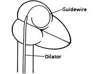
Figure 2.
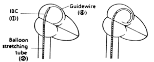
Placement of the Inoue balloon into the correct position in the left atrium.
Figure 3.
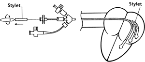
Manipulation of the dilating balloon through the mitral valve.
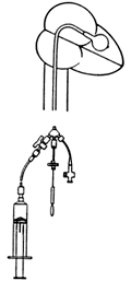
The Inoue balloon inflates in stages and is calibrated, typically from 24 to 28 mm in diameter. With injection of a small amount of diluted contrast material the distal tip of the balloon inflates and is then retracted into the valve orifice (Figure 4).
Figure 4. Inflation of the distal end of the balloon followed by gentle pull-back into the mitral orifice
The balloon is then inflated further, which inflates the proximal portion of the balloon, centering the balloon in the mitral valve orifice. Subsequent inflation stretches the middle of the balloon and splits the commissures of the valve Figure 5).
Figure 5. Angiographic Images Obtained During Mitral Valvuloplasty.
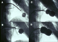
From Feldman T, Herrmann HC, Inoue K: Cath and Cardiovasc Diag. 1994;2:26-34, with permission.
The balloon is rapidly deflated and the valve is re-evaluated hemodynamically and with cardiac echo during the procedure to see if leaflet pliability has improved without production of increased mitral regurgitation. In the absence of new and/or significant mitral regurgitation, the balloon is gradually inflated to larger and larger diameters until the target diameter is achieved. The balloon is then withdrawn and the patient returned to the recovery room. In most instances, the patient leaves the hospital the following day and returns to full activity within three days. Full recuperation for patients undergoing surgical mitral commissurotomy takes, by comparison, six weeks.
Mintral Commissurotomy v. Percutaneous Valvuloplasty
Several randomized trials have been conducted, under the direction of Dr. Zoltan Turi (Harper Hospital, Detroit, Michigan), comparing percutaneous mitral valvuloplasty to both closed and open commissurotomy. These trials showed a very low mortality rate using either surgical or percutaneous techniques. At three-year follow-up, 57% of the surgical patients were functional Class I, while 72% of the percutaneous patients were functional Class I. Additionally, the valve gradient was lower in the valvuloplasty patients at three years and the valve area was increased in the valvuloplasty patients compared to the surgical patients (both desirable outcomes). Mitral regurgitation was similar in both groups.
Other groups have demonstrated that mitral valvuloplasty was successfully carried out in 96% of severe pulmonary hypertensive patients with no reported deaths. Patients with advanced echo scores (10-16), however, do not seem to fare as well and probably should be referred for valve replacement. The mortality in this group was 4% acutely and 9% at one year. The three-year survival was only 42% compared to 77% in the Inoue registry.
Conclusions
Patients with mitral stenosis without significant mitral regurgitation, having an echo score of less than or equal to eight and normal left ventricular function, can be treated with percutaneous mitral valvuloplasty safely with equivalent or better outcomes compared to open or closed mitral commissurotomy. Yet, perhaps because this procedure is relatively new, it is employed considerably less frequently than is indicated.
Since the duration of the procedure is 70 minutes or less in experienced centers and the hemodynamic results are excellent, percutaneous mitral valvuloplasty appears to be the preferred approach.
I hope any Cyberounds® colleagues with experiences --- pro and con --- related to this treatment option will post their comments in the Q&A area to the left of the conference. Questions are also welcome.
In the coming months, I am interested in discussing issues of coronary artery disease reversal, using intensive risk factor interventions, recent advances in percutaneous coronary revascularization and an in-depth review of congestive heart failure. If there's a topic you'd like to see covered or an issue we might raise and discuss here, let me know by posting in the Discussion forum.