Course Authors
Stephen J. Pandol M.D.
Release Date: 08/08/2006
Upon completion of this Cyberounds®, you should be able to:
Diagnose and determine the causative factors in a case of acute pancreatitis
Estimate the severity and level of care needed for a case of acute pancreatitis
Identify those patients with acute pancreatitis who should receive antibiotics
Discuss the issues related to nutrition in acute pancreatitis
Identify patients with acute pancreatitis who require endoscopic and/or surgical treatments.
Acute pancreatitis is a disorder primarily of the exocrine pancreas involving varying degrees of acute inflammation and parenchymal injury of the gland.(1) In its most severe forms, there is a widespread inflammatory response involving organs both adjacent and distant from the pancreas; and the parenchymal injury is manifest as necrosis of pancreatic tissue.
The clinical syndrome resulting from an episode of acute pancreatitis includes the rapid onset of abdominal pain with variable abdominal findings ranging from mild tenderness to peritoneal signs. The disease is often accompanied by vomiting, fever, tachycardia, leukocytosis and increased levels of pancreatic digestive enzymes in the blood and urine. In severe cases, dysfunction is seen in other organ systems (i.e., pulmonary, renal, cardiovascular, hepatic, central nervous system, adipose tissue).
In its simplest classification,(2) acute pancreatitis can be divided into mild and severe:
- Mild disease: minimal organ dysfunction and an uneventful recovery, absent the described features of severe acute pancreatitis;
- Severe disease: multi-organ failure and/or local complications such as necrosis, abscess or pseudocyst.
Epidemiology and Incidence
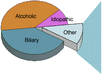
Epidemiological studies demonstrate that approximately 70% of the cases of acute pancreatitis are related to biliary stone disease or alcohol abuse.(1),(2),(3) Other etiologies account for another 20%, and about 10% of cases are idiopathic despite extensive investigation. Although there are many potential etiologies for acute pancreatitis, only a few of the etiologies lead to chronic pancreatitis (i.e., alcohol abuse, genetic disorders and hyperlipidemia). The mechanisms underlying the transition from acute pancreatitis to chronic pancreatitis are presently unknown.
The reported incidence for acute pancreatitis varies widely (from 5-73 per 100,000) and is only an approximation.(1) The reason is that pancreatitis is missed in many patients because the diagnosis is presumptive and based on clinical assessment, laboratory and imaging tests, all having limitations in their sensitivity and specificity. Also, many patients may not come to medical attention because of mild disease and/or limited access to medical care.
There is a slightly greater incidence of acute pancreatitis in males compared to females reported from some countries.(1) Furthermore, in reports where precipitating factors are considered, alcohol abuse is more commonly associated with male cases than female cases, whereas gallstone disease is more commonly associated with female cases than male cases. Thus, the total incidence and the gender distribution of cases of acute pancreatitis in different areas of the world are greatly influenced by the incidence of alcohol abuse and gallstone disease.
Mechanism of Disease
As indicated above, the clinical disorders associated with the majority of cases are alcohol abuse and biliary stone disease. How these two associations are involved in the causation and/or initiation of pancreatitis is not established. Significant progress, however, has been made in identifying key steps that mediate the major pathobiological processes (i.e., inflammation/edema and necrosis) that occur in pancreatitis using experimental animal models of pancreatitis.(4),(5) These models allow investigations into the molecular pathways that mediate the major pathobiological processes. Such investigations are leading to identification of molecular targets for therapy. These include signals in the inflammatory pathway and pathways leading to necrosis. Because these targets are likely utilized in the mechanism of most forms of acute pancreatitis, agents that address these steps will likely have therapeutic benefit.
The major pathobiological processes of acute pancreatitis are influenced by the participation of unique pancreatic intracellular and extracellular events/factors.(3) Examples of intracellular events are activation of digestive enzymes and inhibition of secretion that occur during pancreatitis. Examples of extracellular factors include vascular (i.e., ischemia) and neural participation in pancreatitis. Treatments that modulate these events/factors during pancreatitis may also lead to beneficial outcomes. Such treatments may include protease inhibitors; and blockade of neurotransmitters.
As is evident below, there are presently no specific therapies that include agents that address the pathobiological processes listed above. This is because the information about the mechanisms is recent and rapidly developing. Application of these findings to treatments should now start to advance.
Diagnostic Criteria
A patient with acute pancreatitis most commonly comes to medical attention because of severe and continuous epigastric abdominal pain sometimes radiating to the back. The pain is often associated with nausea and vomiting. An examination of the patient usually reveals epigastric or generalized abdominal tenderness and guarding. The patient may also have tachycardia, hypotension and evidence of volume depletion. Rarely, patients with acute pancreatitis will have periumbilical bruising (Cullen's sign) or flank bruising (Grey Turner's sign) which suggest there is hemorrhage in the pancreas with the pancreatitits (i.e., hemorrhagic pancreatitis).
In a patient with symptoms and signs suggestive of acute pancreatitis, diagnostic tests including measures of serum enzymes (amylase and lipase) and pancreatic imaging studies (i.e., ultrasound, CT) are needed to establish the diagnosis. Table 1 presents an overview of the diagnostic tests.

Table 1. Standard Diagnostic Tests.
| Test | Sensitivity | Specificity | Comment |
|---|---|---|---|
| Serum enzymes | high | moderate | > 3x normal increases specificity |
| Ultrasound | moderate | high | Best for gallstones |
| CT | moderate | high | Detects calcifications, fluid collections |
| CT with pancreatic protocol and IV contrast | moderate | high | Detects necrosis |
Adapted from (3), www.gastroslides.org
When applied to the diagnosis of acute pancreatitis, the following characteristics of the serum tests should be considered:
- Of the commonly measured serum enzymes, lipase is more specific and sensitive and is increased for a longer time compared to serum amylase. Both amylase and lipase can be falsely elevated and both depend on renal clearance. What distinguishes lipase from amylase is that lipase is reabsorbed in the renal tubules, resulting in a more prolonged increased level in the blood. Thus, lipase has a longer half-life (6.9-13.7 hours) than amylase (about 2 hours).
- Serum amylase and lipase usually rise early in the presentation and amylase can be cleared quickly because of its shorter half-life and renal clearance.
- A study comparing 39 patients with acute pancreatitis to 127 controls with abdominal pain showed that the specificity of amylase was 88% while that of lipase was 99%.(1) Other causes for increased serum amylase and lipase are listed in Tables 2A and 2B.
- An increase in serum lipase greater than three times the upper limits of normal almost always indicates a diagnosis of acute pancreatitis.
- Occasionally, alcoholics may have an elevated amylase that is of salivary origin and cannot be distinguished with the more common assays of amylase.
- Serum amylase levels may be normal in pancreatitis related to alcohol abuse, as well as in cases related to hypertriglyceridemia. With respect to alcohol abuse, the normal amylase levels are due in part to the fact that alcohol suppresses amylase synthesis and in part from loss of parenchymal cells containing amylase secondary to chronic injury from alcohol (i.e., chronic pancreatitis).
- Gallstone pancreatitis tends to have greater elevations of amylase and lipase than do other etiologies.
- Since pancreatitis severity does not correlate with measured levels of amylase and lipase, these assays have not been included in the prognostic scoring systems described later. A variety of other causes of increased serum amylase and lipase are listed in the tables that follow.
Table 2a. Causes of Increased Serum Amylase.
| GI Related | Non-GI Related |
|---|---|
| Pancreatic Tumors | Ectopic pregnancy |
| Cholecystitis | Salpingitis |
| Peptic Ulcer Disease | Ovarian cyst |
| Bowel ischemic, perforation, infarction or obstruction | Cystadenocarcinoma of the Ovary |
| Appendicitis | Mumps |
| Sphincter of Oddi Spasm | Diabetic ketoacidosis |
| Morphine administration | Carcinoma of the lung |
| Endoscopic procedures | Renal failure |
Table 2b. Causes of Increased Serum Lipase.
| GI Related | Non-GI Related |
|---|---|
| Acute cholecystitis | Diabetic ketoacidosis |
| Bowel obstruction or infarction | HIV |
| Duodenal ulceration | Macrolipasemia |
| Pancreatic calculus | Idiopathic |
| Pancreatic tumors | Drugs |
| Post-ERCP/trauma |
Evaluation for Etiologic Factors
Figure 1 provides an overview of various factors associated with the development of acute pancreatitis. Although the mechanisms by which these factors influence the development of pancreatitis are not entirely known, it is important to identify the associated factors in patients with acute pancreatitis because the removal of the factor(s) may decrease the risk of recurrent episodes of acute pancreatitis. Some examples include cholecystectomy in patients with cholelithiasis; counseling for alcohol abuse; correction of metabolic causes such as hypertryglyceridemia and hypercalcemia; identification and stopping of causative medications (Table 3); and treatment with corticosteroids for autoimmune pancreatitis. Also, of significant importance in the older patient (i.e., 50 years of age or older), identification of an early pancreatic cancer as a cause of pancreatitis may prevent mortality from the cancer.
The etiologic factor for a patient with an episode of acute pancreatitis or recurrent pancreatitis may not be identified during the initial evaluation or even after a recurrent episode. In this case, invasive imaging techniques (i.e., endoscopic ultrasound with biopsy and endoscopic retrograde cholagiopancreatography) should be considered to evaluate for pancreatic tumors and pancreatic duct anomalies. In addition, there should be consideration of disorders such as autoimmune pancreatitis and genetic causes (i.e., cystic fibrosis and hereditary pancreatitis). Autoimmune pancreatitis can be diagnosed by imaging and biochemical testing, while the genetic etiologies require formal genetic testing. For more detailed approaches to these uncommon etiologies, see the articles by Whitcomb and Okasaki et al.(6),(7)
Figure 1. Etiologies of Acute Pancreatitis.
|
|
Adapted from (3), www.gastroslides.org
Table 3. Drug Induced Acute Pancreatitis.
| Common | Uncommon | Rare |
|---|---|---|
| asparaginase | ACE inhibitors | carbamazepine |
| azathioprine | acetaminophen | corticosterioids |
| didanosine (ddi) | 5-amino ASA | metronidazole |
| 6 mercaptopurine | furosemide | minocycline |
| pentamidine | sulfasalazine | nitrofurantoin |
| valproate | thiazides | tetracycline |
Adapted from (3), www.gastroslides.org
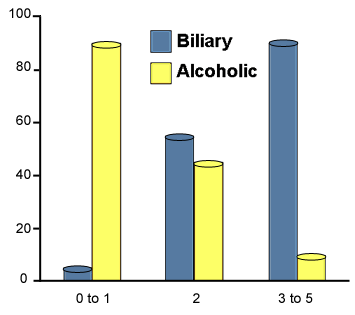
As illustrated above, alcohol-induced and gallstone- or biliary-induced acute pancreatitis are the two most common etiologic factors for acute pancreatitis. In addition to obtaining history related to alcohol abuse and imaging studies to determine the presence of gallstone disease, Figure 2 below shows the predictive values of age, sex and serum tests in distinguishing between alcoholic and biliary pancreatitis.
Figure 2. Predictors of Etiology.
# Predictors
|
|
Adapted from (3), www.gastroslides.org
Measures of Severity/Predictors of Outcome
Early staging of severity is critical for identifying patients who require intensive monitoring and support. Several systems have been developed and are described in more detail below with links to Websites.
Ranson's Signs, APACHE II (Acute Physiology And Chronic Health Evaluation) and Balthazar's CT Severity Index (CTSI) are commonly used criteria to assess severity and outcome in pancreatitis patients.(1),(2),(3) APACHE II is a measure of disease severity and outcome prediction using several clinical and laboratory observations. Severe pancreatitis, with a potential for significant morbidity and mortality, occurs when one or more of the following are present (from 1992 Atlanta Classification System):
- Ranson's criteria > 3
- APACHE II score > 8
- Necrosis on contrast enhanced CT scan (CTSI)
- Organ failure and/or local pancreatic complications
- Pancreatic abcess
- Pancreatic pseudocyst.
These patients require intensive care unit (ICU) monitoring and treatment of local and systemic complications:
Calculations and Mortality Prediction Using Ranson's Criteria
Table 4 and Figure 3, as well as the Website below, provide the necessary information for calculation of severity using Ranson's criteria.
Table 4. Ranson's Criteria of Severity.
| At admission | During initial 48 hours |
|---|---|
|
|
Adapted from (3), www.gastroslides.org
Figure 3. Mortality Related to Ranson's Criteria.
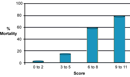
The majority of patients with severe disease have from 3 to 5 criteria.
Adapted from (3), www.gastroslides.org
Calculations and Mortality Prediction for APACHE
Go to the following Website for the calculation of the APACHE II score for severity: www.sfar.org/scores2/apache22.html
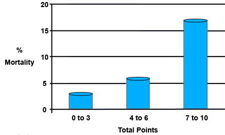
Calculations and Mortality Prediction Using CTSI
This scoring system (Table 5 and Figure 4) uses several CT observations including edema and fluid collections. Importantly, the estimates of the amount of necrosis of the gland, as determined by measuring the portion of the gland that perfuses with contrast injection, is highly predictive of necrosis and poor outcome. Stated simply, greater amounts of estimated necrosis are associated with greater rates of extrapancreatic organ failure and pancreatic infection occurring during the course of the episode.
Table 5. CT Severity Index.
| CT finding | Points |
|---|---|
| Gland enlarged | 1 |
| Peripancreatic inflammation | 2 |
| One fluid collection | 3 |
| Multiple fluid collections | 4 |
| Necrosis <30% | 2 |
| Necrosis 30 - 50% | 4 |
| Necrosis >50% | 6 |
Adapted from (3), www.gastroslides.org
A Practical Approach for Predicting Severity
A practical approach to the measurement of severity is to monitor early clinical indicators of severity (Table 6) and perform a contrast-enhanced CT to determine the CTSI. Finally, although not considered in the measurement systems described above, a measurement of the blood C-reactive protein (CRP) is an easy assay with predictive value, as illustrated in Figure 5.
It should be noted that CRP rises higher and persists longer with severe pancreatitis.(8) Peak values of CRP of 150-200 or more on days 2-4 or greater than 120 at the end of the 1st week are predictors of severe pancreatitis. The accuracy of using CRP for measurement of severity is similar to Ranson's scoring system.
Table 6. Early Indicators of Severity.
|
Adapted from (3), www.gastroslides.org
Figure 5. C-reactive Protein (CRP) Levels in Acute Pancreatitis.
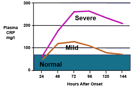
Adapted from (3), www.gastroslides.org
Management of Acute Pancreatitis
Supportive Care
The management of acute pancreatitis is largely supportive (Table 7). Patients with mild pancreatitis can be monitored and managed in the non-ICU setting. However, patients with more severe pancreatitis (i.e., presence of early indicators of severity, CRP greater than 150 and/or greater than 30% necrosis of the gland, as deterimined by contrast-enhances CT) require monitoring and treatment in the ICU. Monitoring should include measurements of neurological status, vital signs, arterial oxygen saturation, renal function, abdominal physical findings and serum electrolyte (including Ca 2+) concentrations. The graphic below lists general supportive care.
Table 7. Treatment for Acute Pancreatitis.
| Supportive Care | Other Treatments |
|---|---|
|
|
Adapted from (3), www.gastroslides.org
Other Treatments
Although acid suppression is frequently used, its benefits are unclear. Nasogastric suction has not been shown to shorten the course of acute pancreatitis but should be used in patients with nausea and vomiting to relieve the discomfort.
In patients with a severe or a worsening clinical course of pancreatitis, especially with signs of biliary sepsis (i.e., fever, increasing WBC, bilirubin and alkaline phosphatase levels), urgent imaging with abdominal ultrasound or CT scan should be performed. Serial CT scans should also be considered to follow the evolution of necrosis. MRI can also evaluate for pancreatic necrosis for those with renal insufficiency or contrast allergy.
If the clinical course and imaging studies demonstrate biliary obstruction and sepsis from stones, an urgent ERCP (endoscopic retrograde cholangiopancreatography), combined with sphincterotomy, should be considered for stone removal.
In patients who have signs of pancreatic infection (infected necrosis, pseudocyst or abcess) by clinical course and CT imaging, the suspicious pancreatic lesion should be sampled to test for infection. For lesions found to be infected, debridement and large volume lavage by interventional radiology and/or surgery must be performed. In addition, antibiotic therapy is necessary. In patients who develop evidence of sepsis while taking antibiotics to prevent infection, one must consider superinfection with Candida and/or Staphylococcus. For these patients, changing antibiotic coverage and/or adding fungal coverage may be necessary.
Antibiotics for the Prevention and Treatment of Infections
As discussed above, antibiotics are used to both prevent infections as well as to treat established infections during pancreatitis. For patients with severe pancreatitis, as determined by prognostic indicators, one should institute preventive antibiotic therapy. However, this is a controversial topic.
A recent meta-analysis showed that there was a benefit in using prophylactic antibiotics in reducing mortality but the advantage was limited to patients with severe acute pancreatitis who received broad-spectrum antibiotics capable of achieving therapeutic pancreatic tissue levels.(9) The largest Randomized Control Trial (ciprofloxacin + metronidazole vs. placebo) involving 114 patients showed no benefit.(10) Furthermore, the development of resistance and or superinfections with staph or fungal infections can worsen morbidity and mortality. Thus, this area of management is fraught with controversy. Antibiotics and antibiotic combinations that are used in preventive strategies include imipenim alone; cefurozime alone; ceftazidime, amikacin and metronidazole; and ciprofloxacin and metronidazole.
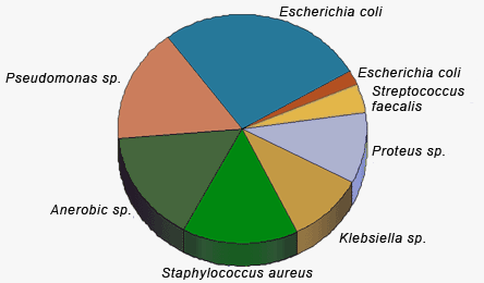
Also, as indicated earlier, antibiotics or changes in antibiotic therapy are necessary for the treatment of biliary sepsis with gallstone pancreatitis and infected pseudocysts or pancreatic necrosis. Figure 6 provides information on the relative contribution of different microorganisms to infected pancreatic necrosis.
Pain Management
Abdominal pain is frequently the dominant symptom in acute pancreatitis. In severe pancreatitis, uncontrolled pain can contribute to the hemodynamic instability. Adequate pain control requires the use of intravenous narcotics. In the past, meperidine was favored over morphine for analgesia in pancreatitis because human studies showed that morphine caused increases in sphincter of Oddi pressure. Despite these data there is no clinical evidence to suggest that morphine can aggravate or cause pancreatitis or cholecystitis. Because repeated doses of meperidine can lead to accumulation of the metabolite, normeperidine, which causes neuromuscular irritation and rarely seizures, its use has diminished.
Appropriate agents to use for pain management are hydromorphone (Dilaudid), fentanyl and morphine. The initial dose of morphine is usually 2 mg given parenterally every 4 hours. Recommended initial doses and frequencies of administration are provided in Table 8.
Table 8. Pain Medications: Dosage and Frequency.
| Medication | Initial Dose | Frequency | Usual Dose Range |
|---|---|---|---|
| Hydromorphone | 0.2 mg | 4 hours | 0.2-1.5 mg |
| Fentanyl | 25 μg | 4 hours | 25-100 μg |
| Morphine | 2 mg | 4 hours | 2-10 mg |
All narcotics can cause depression of respiratory and cardiovascular functions, which can be exacerbated by other agents used in these patients such as calcium channel blockers and beta-adrenergic antagonists.
Nutritional Management
Patients with mild acute pancreatitis should remain NPO until the pain resolves, bowel sounds become normal and the patient is hungry. The diet should advance slowly as the patient tolerates food -- so long as the pain, nausea and vomiting remain absent and the patient continues to have a desire to eat. There is a risk of relapse of the acute pancreatitis if the patient is given oral feedings too soon or the diet is advanced too rapidly.
In those patients with severe pancreatitis and whose symptoms are not resolving within 5 days, caregivers should consider implementation of enteral feeding or TPN. There is accumulating evidence that enteral feeding has advantages over TPN in terms of both cost and outcome and it is becoming the preferred nutritional support strategy.(11) For enteral feeding, the current practice is to deliver nutrients into the jejunum through feeding tubes.
Current clinical practice dictates that patients with pancreatic ascites, enlarging pseudocysts or peripancreatic fluid collections should remain NPO and receive nutrition by TPN until these problems resolve. However, as discussed in the previous paragraph, there is an evolving understanding of the benefit/risk of enteral feeding in pancreatitis. Thus, it is possible that feeding practice will also change in this subset of patients.
Management of Complications
Complications of acute pancreatitis, in addition to the infections previously described, include fluid collections (pseudocysts, effusions and ascites) and bleeding.
Pseudocysts are collections of pancreatic fluid contained by granulation tissue and fibrosis and not by an epithelial lined structure. Thus, the name pseudocyst. The fluid contains high concentrations of pancreatic enzymes. Pseudocysts are common and appear in up to 10% of patients with acute pancreatitis. They may be either asymptomatic or present with upper abdominal pain, early satiety and or nausea/vomiting. These symptoms are in part due to partial gastric obstruction resulting from the pseudocyst. Complications of pseudocysts include rupture and leakage of pancreatic fluid into the peritoneal cavity (i.e., pancreatic ascites) and hemorrhage.
Most pseudocysts resolve spontaneously. Thus, the current recommendation, as outlined in Figure 7, is to monitor until resolution and to consider drainage when the patient has symptoms; when there is evidence of infection; or when the pseudocyst continues to enlarge. There are three major types of drainage techniques. Both a surgical and an endoscopic technique create a cyst-gastrostomy for the pseudocyst to drain into the stomach. An interventional radiologic approach consists of inserting a drainage catheter percutaneous that drains the fluid externally.
Figure 7. Pseudocyst Management.
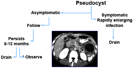
* Large cysts can be safely followed, but are more likely to require drainage.
Adapted from (3), www.gastroslides.org
Pancreatic ascites is a complication of a leaking pseudocyst or disruption of the pancreatic ductal system. The measures of ascites fluid diagnostic for pancreatic ascites are listed in Table 9. Treatment of pancreatic ascites includes decreasing secretion of the pancreas using octreotide and TPN (i.e., decrease stimulation of the pancreas); removal of the ascites fluid by paracentesis; pancreatic duct stenting; and surgical repair.(12)
Table 9. Characteristics of Ascites.
| Protein | *SAAG | Amylase | WBC's | |
|---|---|---|---|---|
| Peritoneal burn | High | Low | Moderate | High |
| Pancreatic | High | Usually low | High | Moderate |
| Cirrhotic | Variable | High | Low | Low |
* SAAG: serum-ascites albumin gradient
Adapted from (3), www.gastroslides.org
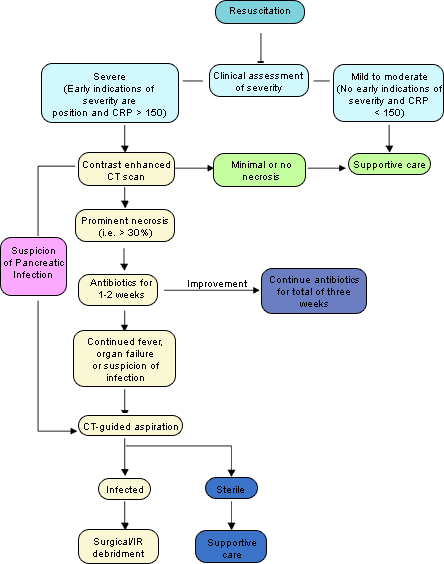
Algorithm of Management
The algorithm below summarizes the major issues in management:
Summary
Acute pancreatitis is a disorder with a wide spectrum of manifestations. The key to providing optimal therapies for these patients is recognizing the severity of pancreatitis as well as the cause. Issues of monitoring, supportive care, anticipating complications for rapid treatment and preventing recurrences all flow from defining the severity and cause as early as possible in the course of an episode.