Course Authors
Nicole D. Simpson, M.D., Tommy Yen, M.D., and Aijaz Ahmed, M.D.
From the Department of Medicine, Division of Gastroenterology, Stanford University School of Medicine, Stanford, California; Division of General Internal Medicine and Geriatrics, Veteran's Adminstration Medical Center, San Diego, California; and University of California San Diego School of Medicine, San Diego, California.
Drs. Simpson, Yen and Ahmed report no commercial conflicts of interest.
Estimated course time: 1 hour(s).

Albert Einstein College of Medicine – Montefiore Medical Center designates this enduring material activity for a maximum of 1.0 AMA PRA Category 1 Credit(s)™. Physicians should claim only the credit commensurate with the extent of their participation in the activity.
In support of improving patient care, this activity has been planned and implemented by Albert Einstein College of Medicine-Montefiore Medical Center and InterMDnet. Albert Einstein College of Medicine – Montefiore Medical Center is jointly accredited by the Accreditation Council for Continuing Medical Education (ACCME), the Accreditation Council for Pharmacy Education (ACPE), and the American Nurses Credentialing Center (ANCC), to provide continuing education for the healthcare team.
Upon completion of this Cyberounds®, you should be able to:
List risk factors for gallstone formation
Discuss complications precipitated by gallstones
Discuss benefits of timely consultations especially in terms of improving clinical outcome.
Prompt recognition of the type and severity of complications precipitated by gallstones is crucial for a successful outcome. Timely consultative input from surgical, radiology and gastroenterology services can minimize the incidence of poor clinical result.
Composition of Gallstones
Based on their chemical composition, gallstones can be divided into three types: cholesterol, pigmented or mixed. Up to 90% of gallstones have cholesterol as their major constituent and 10% of gallstones are pigmented. The black-pigmented stones are associated with chronic hemolytic disease, while brown-pigmented stones are a consequence of biliary tract infections.
Mechanism of Gallstone Formation
The underlying pathogenetic mechanisms that result in gallstone formation include supersaturation of bile constituents, bile stasis and presence of nucleation factors such as mucin, glycoproteins and calcium. The predisposing risk factors for gallstone formation are listed in Table 1:

Table 1. Risk Factors for Cholesterol Gallstone Formation.
| Table Headline |
|---|
|
Because of the overlapping symptom spectrum, gallstone complications may result in misdiagnosis.(1),(2)The management is variable and depends on the extent, severity and type of gallstone-induced complication. For example, a management plan for biliary colic may include conservative follow-up or elective cholecystectomy. On the other hand, an urgent laparoscopic cholecystectomy within 24 to 48 hours is indicated in a hemodynamically stable patient with acute cholecystitis.
Clinical Manifestations of Gallstone Complications
Up to one-third of patients with gallstones are symptomatic.(3) Biliary colic is the most common clinical presentation and is noted in 70% to 80% of symptomatic patients with gallstones.(4) Typically, biliary colic is characterized by severe, episodic pain located in the epigastrium or less frequently in the right upper quadrant. The term biliary colic is a misnomer because the associated pain is steady and not colicky. Intolerance to dietary fat is not a feature of biliary colic. A large meal may precipitate an attack of biliary colic. However, more often there are no precipitating events.
Biliary colic occurs more commonly at night, the pain developing suddenly and increasing in intensity over a 15-20 minute period to a steady plateau which can last for several hours. The pain may radiate to the interscapular region or to the right shoulder. Nausea, vomiting and sweating are not uncommon. Pain may subside slowly or decrease in intensity to a residual persistent abdominal pain. Pain lasting more than three hours is usually associated with the development of cholecystitis. The time period between biliary colic attacks is variable and may be weeks, months or even years.
Acute cholecystitis is precipitated by gallstones in 90% of cases.(5) The remaining 10% cases of acute cholecystitis are acalculous or infectious. Acute cholecystitis can result in local inflammation (e.g., right upper quadrant mass, tenderness) and systemic toxicity (e.g., fever, leukocytosis). Typically, the pain of acute cholecystitis lasts longer than three hours and with time may shift from the epigastrium to the right upper quadrant. A pain-free period lasting several hours between epigastric and right upper quadrant pain may be noted. Any movement including cough or sneeze aggravates the pain. Vomiting and low-grade fever is common.
Elderly patients may not present with pain or fever, and localized tenderness may be the only symptom. Murphy's sign, the sudden arrest of breathing during inspiration secondary to pain, is elicited by deep palpation of the right upper quadrant. Jaundice develops in 15% of patients with acute cholecystitis, even in the absence of choledocholithiasis (the presence of gallstones in the common bile duct).(6)
During an episode of acute cholecystitis, an impacted stone within the cystic duct triggers an inflammatory response that produces edema and compression of the common bile duct (CBD), common hepatic duct or both.(7) Therefore, patients can develop jaundice in the absence of common bile duct or common hepatic duct stones (Mirizzi's syndrome).
Prolonged cystic duct obstruction can result in gallbladder distension with clear mucoid fluid, a condition known as hydrops of the gallbladder. Less frequently, a large gallstone will erode through the gallbladder wall into the duodenum. Subsequently, the stone may lodge at the terminal ileum with small bowel obstruction or the duodenal bulb/pylorus causing gastric outlet obstruction (Bouveret's syndrome). Recurrent attacks of biliary colic or acute cholecystitis can result in chronic cholecystitis.
Acute acalculous cholecystitis affects predominantly males who are over 50 years in age. It is more frequently noted in the setting of major surgery, critical illness, extensive trauma, burn-related injury or in patients on total parenteral nutrition. Bile inspissation or sludge formation may be the underlying pathogenetic mechanism. Acalculous cholecystitis is associated with a higher mortality rate than calculous cholecystitis.
Rarely, acute cholecystitis is caused by specific infections, such as salmonellosis. Cytomegalovirus and cryptosporidia have been reported to precipitate cholecystitis and cholangitis in severely immunocompromised patients, such as those with acquired immunodeficiency syndrome or after bone marrow transplantation.
Diagnosis of Gallstone-induced Complications
A self-limited episode of biliary colic without progression to acute cholangitis usually does not result in changes in hematological and biochemical tests.(8) In acute cholecystitis, leukocytosis with an increase in immature leukocytes can be noted. Transient pancreatic duct obstruction may result in elevated serum amylase level. Edema and inflammation of the gallbladder can impinge on the common bile duct and result in mild elevation of the serum aminotransferases and alkaline phosphatase. In addition, hyperbilirubinemia can be associated with the enzyme elevation. Aminotransferase elevation can be noted within hours following the onset of the abdominal pain. Gallstone-induced pancreatitis is associated with an elevation in alanine aminotransferase (ALT) of greater than 2.5 times above normal.
Ultrasonography is a reliable diagnostic tool to document stones within the gallbladder.(9) It has greater than 95% sensitivity and specificity in detecting gallstones greater than 2 mm in diameter. Ultrasonographic study of the gallbladder should be preceded by a fast of at least 8 hours because gallstone detection rate improves when the gallbladder is distended and bile-filled. Although small gallstones (<2 mm in diameter) may be difficult to visualize, sludge can precipitate biliary colic, acute cholecystitis or pancreatitis and is regarded as part of the spectrum of gallstone disease.
Ultrasonography less accurately detects choledocholithiasis and may fail to identify up to 50% of stones lodged in the bile ducts. Ultrasound scans have 76% sensitivity in detecting dilatation of intrahepatic or extrahepatic bile ducts, a finding highly suggestive of distal obstruction. Ultrasound is routinely used in the diagnosis of acute cholecystitis by documenting pericholecystic fluid in the absence of ascites, gallbladder wall thickening greater than 4 mm in the absence of hypoalbuminemia and sonographic Murphy's sign.
Endoscopic retrograde cholangiopancreatography (ERCP) is the gold standard for the diagnosis of choledocholithiasis. ERCP has 95% sensitivity and 95% specificity to detect bile duct stones and has the added benefit of therapeutic options. When there is a need to visualize the biliary system directly for diagnostic or therapeutic reasons, an ERCP can be employed.
The recent improvement in the processing of CT and MRI data into a three dimensional image offers diagnostic accuracy that is comparable to ERCP. But these modalities do not provide any therapeutic options. Therefore, noninvasive CT and MRI may be used to exclude choledocholithiasis in patients with low probability of biliary ductal stones, while ERCP can be used in those with high suspicion of choledocholithiasis.
Hepatobiliary scintigraphy can be employed with a high degree of sensitivity and specificity to confirm or exclude the diagnosis of acute cholecystitis.(10) After a two to four hour fast, an intravenous injection of a 99mTc-labeled iminodiacetic acid derivative (IDA agent) is instituted following the fast. The IDA agent is excreted into the bile ducts. Serial imaging is conducted.
In a normal study, images of the gallbladder, common bile duct and small bowel appear by 30 to 45 minutes. Failure to image the gallbladder by 90 minutes, despite adequate views of the liver, CBD and small bowel, strongly suggests acute obstructive disease. A normal 99mTc-IDA scan reliably excludes the diagnosis of acute cholecystitis in a patient with abdominal pain.
An abnormal or 'positive' 99mTc-IDA scan constitutes nonvisualization of the gallbladder with documentation of preserved excretion into the common bile duct and small bowel. Patients with acalculous cholecystitis may have a false negative study. False positive results can be noted in patients with inadequate fasting, prolonged fasting states (TPN use), chronic alcoholism and chronic cholecystitis that result in gallbladder stasis and poor filling. Follow-up scans at 4 or more hours decreases the false positive rate and increases the sensitivity and specificity to 97% and 90%, respectively.
Management of Gallstone-induced Complications
The management of gallstone-induced complications varies with the specific type of clinical presentation.(11)
Asymptomatic Gallstones
Based on natural history studies, 60% to 80% of all gallstones are asymptomatic. The risk of developing a symptomatic gallstone-induced complication is relatively low. Therefore, patients with silent or incidental stones should be observed and managed conservatively.
The potential exceptions to this recommendation are specific groups thought to be at higher risk for the development of gallbladder carcinoma. These patients include asymptomatic individuals with a generally calcified (porcelain) gallbladder, gallstones greater than 2.5 cm in size, gallbladder polyp greater than 10 mm in diameter, and certain Native Pima Indians. Because the natural history of gallstones in diabetics follows the same pattern as in nondiabetics, prophylactic cholecystectomy is not recommended in diabetics with asymptomatic gallstones.
Biliary Colic
The risk of recurrent attack is high subsequent to an initial episode of biliary colic. Studies that followed symptomatic gallstone patients indicate a 38% to 50% per year incidence of recurrent biliary pain. Up to 90% of complications, including acute cholecystitis, cholangitis or pancreatitis, are preceded by attacks of biliary colic. There is a 1 to 2% per year risk of developing biliary complications after an initial attack of biliary colic.
As many as 30% of patients who are followed long-term after an attack of biliary colic remain asymptomatic. The decision to perform a laparoscopic cholecystectomy should be individualized based on surgical candidacy of the patient; a healthy patient with no surgical contraindications should be recommended to have laparoscopic cholecystectomy.
Acute Cholecystitis
An urgent laparoscopic cholecystectomy should be performed once the diagnosis of acute cholecystitis is made in a hemodynamically stable patient. The optimal time to perform a laparoscopic cholecystectomy is within 24 to 48 hours following presentation.
Choledocholithiasis
Severe acute cholangitis or acute biliary pancreatitis precipitated by gallstones should be managed by an emergent therapeutic ERCP for biliary decompression. In a patient with choledocholithiasis, a variety of therapeutic interventions can be instituted. In general, patients with jaundice and documented biliary ductal dilatation should be promptly scheduled for a preoperative ERCP examination with possible sphincterotomy and stone extraction. The patient can then undergo a routine laparoscopic cholecystectomy within 1 or 2 days. On the other hand, if the liver enzymes are mildly abnormal and there is a low suspicion for common bile duct stones, many physicians proceed directly with laparoscopic surgery.
An intraoperative cholangiography can be performed to rule out choledocholithiasis. There are two options in patients suspected to have choledocholithiasis during the intraoperative cholangiogram. First, depending on the surgeon's experience, intraoperative laparoscopic stone removal may be attempted through the cystic duct with balloon catheters, basket retrieval devices, choledochoscopes or irrigation techniques. Second, the surgeon may elect to convert to an open common bile duct exploration or to arrange for a postoperative ERCP.
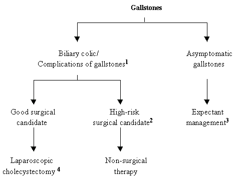
Elderly patients, who present with symptomatic common bile duct stones and who are at high risk for surgical complications or patients who decline surgery, may benefit from endoscopic sphincterotomy as primary therapy for cholelithiasis or choledocholithiasis (Figure 1):
Figure 1. Management of Gallstones.
Click to see full sized image
- Emergent therapeutic ERCP in patients with acute gallstone pancreatitis or acute cholangitis.
- Percutaneous cholecystostomy for gall bladder decompression and drainage in acute cholecystitis.
- Prophylactic cholecystectomy in patients with asymptomatic gallstones: Pima Indians; calcified gallbladder, gallbladder polyps >10 mm, and gallstones >2.5 cm.
- Choledocholithiasis: ERCP (preoperative or postoperative) or intraoperative cholangiography.