Course Authors
Walter S. Lesley, M.D.
Release Date: 05/08/2002
Upon completion of this Cyberounds®, you should be able to:
Discuss the utility of vertebroplasty to treat osteoporosis
Identify ideal candidates for vertebroplasty
Discuss the complications of vertebroplasty.
The risk of fracture increases with age in postmenopausal women. Lifetime risk of vertebral fracture from age 50 onward is 16% in white women and 5% in white men. Fracture rates are lower in other racial groups. Moderate or severe vertebral compression deformities are found in 25% of 85-year-old women.
In 1995, osteoporotic fractures accounted for 2.5 million physician visits, 432,000 hospital admissions, 180,000 nursing home admissions and $13.5 billion in direct medical expenses.
Osteoporotic fractures lead to impaired quality of life from pain, disability, loss of activities of daily living, fear of falling, insomnia, embarrassment regarding kyphotic appearance and depression.
The diagnosis of osteoporotic vertebral compression fractures involves a vertebral body height reduction of >15%. They are classified by type, viz., wedge, biconcave and compression. Fifty-nine percent of vertebral compression fractures occur spontaneously, clinically.
Diagnosis
Eighty-four percent of vertebral compression fracture patients have associated pain:
- Pain lasts 2 weeks to 3 months average
- Pain described as deep, intense at or near the level of the involved vertebra, but pain can be referred
- Relieved while supine
- Exacerbated with standing or bending.
Most common locations are T8, T12, L1, L4. Four diagnostic tests are used, viz:
- Plain films
- Fluoroscopy
- MRI
- Nuclear bone scan.
Figure 1. Routine Plain Film Radiography.

Compare with any prior studies.
Figure 2. MRI.
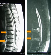
Assess for vertebral body marrow edema.
Exclude spinal canal stenosis, disc, or facet disease
Figure 3. Fluoroscopy Examination.
Establish concordance between painful site clinically and multiple compression fxs on imaging
The Clinical Evaluation
The clinical evaluation includes: pain corresponding to level of the fracture; focal tenderness corresponding to the level on imaging; symptoms present <3 months or recent exacerbation of previous fracture; pain may radiate along flank or anteriorly; exclude radicular pain; exclude cord compression from retrovulsed vertebral body cortex into spinal canal.
Traditional Management
- Bed rest, with variable success
- Complications
- Deep venous thrombosis pneumonia
- May cause further demineralization
- Complications
- Traditional fixation surgery
- High failure rate because of weakened bone foundation
- Analgesics
- Side effects: urinary retention, ileus
- Addiction
A New Treatment Paradigm: Osteoporotic Vertebral Compression Fractures and Vertebroplasty
Figure 4. Vertobroplasty.

Percutaneous injection of methylmethacrylate cement into the affected vertebral body.
IV conscious sedation and local anesthetic
Outpatient procedure
In 1987, the first case of vertebroplasty was reported in France by Galibert et al.(2)
- 50-year-old female with neck pain from cervical (C2) hemangioma.
In 1997, Jensen et al.(3) reported 29 patients with 47 osteoporotic compression fractures treated with vertebroplasty with a 90% pain relief. The following year Deramond et al.(4) treated 80 patients with osteoporotic compression fractures with vertebroplasty with a 90% pain relief reported. In 1999, Cortet et al.(5) performed a prospective study of 20 vertebrae in 16 patients with demonstrated pain relief (p<0.01).
Benefits of Vertebroplasty
- Pain relief
- Improved immediately although some procedure-related soreness for up to 72 hours managed with NSAID
- Improved mobility
- Patient able to stand and walk within 2-24 hours.
Efficacy of Vertebroplasty
In osteoporotic compression fractures, overall, 85-90% of patients experience dramatic or complete relief of pain within 72 hours. In neoplastic compression fracture, 60-70% of patients experience marked reduction in narcotic requirements of complete pain relief.
Vertebroplasty alleviates pain by:
- Stabilization of fracture
- Preventing further collapse of the treated vertebral body
- Debated localized nociceptor denervation from exothermic heat of methylmethacrylate polymerization.
The MMA cement is FDA-approved but use for vertebroplasty is "off-shelf" and, therefore, vertebroplasty is investigational. It is reimbursed in most U.S. locales by Medicare/Medicaid and most insurers.
Vertebroplasty prevents further reduction in height of treated vertebra and does not increase risk of compression fractures at adjacent levels.
Contraindications of vertebroplasty include uncorrected coagulopathy, spinal or systemic infection, and hypersenitivity to the cement.
There is a 90% likelihood of pain relief with vertebroplasty. Complications are minor (3%) and major (1%). The majority of complications are transient and self-limited. Steroid therapy or surgery are rarely required.
Complications
Complications include:
- Hemorrhage
- Infection
- Pulmonary embolism (fat or cement emboli)
- Fracture (lamina or pedical)
- Increased pain
- Death
Figure 5. Complications.

Spinal cord or nerve root injury:
- < 1%
- Direct needle trauma
- Indirect
- Cord compression
- Hematoma
- Cement
- Vascular injury and ischemia
- Cord compression
There have been no cement-related complications in over 1,500 vertebroplasties.(7) There was cord compression (0.4%) in one and radicular pain (1.5%) in three of 258 patients treated with vertebroplasty.(8)
Figures 6 and 7. Vertebroplasty.

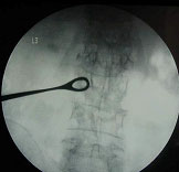
Performed under biplane fluoroscopy

Needle insertion.
Figure 8. Needle Insertion - Unilateral.

Figure 9. Needle Insertion.

Figure 10. Needle Insertion - Bilateral.

Venogram helps visualize epidural and paraspinal draining veins prior to cement injection.
Figure 11. Venogram.

Performed through bone needle.
Injection of small volume of dilute low osmolar nonionic contrast agent.
Figure 12. Vertoplasty.

Cement Mixture
- Polymer powder
- Liquid monomer
- Opacifying agent
- BaSO4 powder
Figure 13. Cement Injection.

Figure 14..

Under biplane fluoroscopy, liquified cement is injected into the vertebral body.
Figure 15..

Complete or bilateral filling of vertebral body is not required for either structural stability or pain relief.(9)
Figure 16. 64 yo Male H/O Leukemia with Acute Low Back Pain.

3 Lumbar compression deformities.
Bone biopsy performed through cannula of bone needle.
Figure 17. Vertebroplasty of L2 and L3.

Patient experienced partial pain relief.
Figure 18. Vertebroplasty of L1.

Patient had complete pain relief.
Biopsy results showed no tumor recurrence.
Post Operative Care
Post operative care includes:
- Dressing at needle site
- Bed rest for 1 hour then advance to ambulation in 2nd hour
- Monitor vital signs
- Monitor neurologic examination
- Discharge at 2 hours
- Gradual increase in activity over 3 days
- Patient may take analgesics prn (decrease dose and frequency)
- Follow-up at clinic in 1 week
- Patient instructed to call if:
- New back pain
- Chest pain or shortness of breath
- Lower extremity weakness
- Fever >100oF
Conclusions
Vertebroplasty is safe for the treatment of pain and disability secondary to osteoporotic compression fractures. There is a low complication rate and high success rate. It is a palliative procedure that does not correct underlying causes of the vertebral fracture. Medical management of osteoporosis or malignancy must be initiated and maintained.