Course Authors
Yusuf Yazici, M.D. and Akgun Ince, M.D.
Dr. Yazici is Attending Physician, Division of Rheumatology, Long Island College Hospital, Brooklyn, NY, and the Hospital for Special Surgery, Weill Medical College of Cornell University, New York, NY and Dr. Ince is Assistant Professor of Internal Medicine, Division of Rheumatology, Saint Louis University School of Medicine, St. Louis, MO. Drs. Yazici and Ince report no commercial conflict of interest.
Estimated course time: 1 hour(s).

Albert Einstein College of Medicine – Montefiore Medical Center designates this enduring material activity for a maximum of 1.0 AMA PRA Category 1 Credit(s)™. Physicians should claim only the credit commensurate with the extent of their participation in the activity.
In support of improving patient care, this activity has been planned and implemented by Albert Einstein College of Medicine-Montefiore Medical Center and InterMDnet. Albert Einstein College of Medicine – Montefiore Medical Center is jointly accredited by the Accreditation Council for Continuing Medical Education (ACCME), the Accreditation Council for Pharmacy Education (ACPE), and the American Nurses Credentialing Center (ANCC), to provide continuing education for the healthcare team.
Upon completion of this Cyberounds®, you should be able to:
Discuss the pathogenesis of osteoarthritis
Describe the clinical presentations of osteoarthritis
Determine, for each patient, the appropriate management of osteoarthritis.
Osteoarthritis (OA) is the most common form of arthritis and is a major cause of morbidity and disability. Already the second leading cause of long-term disability,(1) as the population ages, OA's rising prevalence will further burden health care resources.
Epidemiology
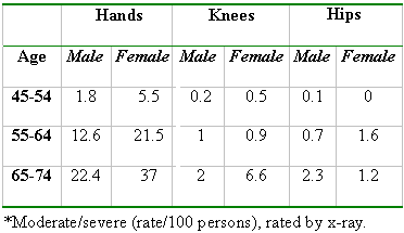
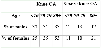
Even though the prevalence of OA can change from population to population, it is a universal problem. Over one-half of all people older than 65 show OA-associated changes in the knees. After age 75, almost everyone has these changes.(2)
Age is the strongest determinant of OA and the prevalence rates for all joints increase with age. Incidence also increases with age but seems to reach a plateau in the seventh decade. The exact mechanism by which age predisposes individuals to OA is not known. Biochemical changes within aging cartilage may render cartilage more susceptible to damage and degeneration but there is not sufficient evidence to support this.(3)
Occupations that subject joints to repetitive trauma may predispose the affected joints to OA. Women are at increased risk of developing OA, especially after menopause.(4) Epidemiologic studies suggest that hormone replacement therapies provide a protective effect from the development of knee and hip OA.(4) Frequency of OA seems to be similar among genders between the ages 45-55.(5) Blacks are more likely to develop knee OA than Whites.
Obesity is more strongly correlated with knee OA than hip OA.(6) Obesity may act by increasing mechanical stress in weight-bearing joints. In addition, obesity may, itself, be a risk factor, especially for hand OA -- obese women are more likely to have OA and as hands do not suffer from mechanical stress, it is postulated that systemic factors may be involved.(3)
Higher rates of hip OA are seen in jobs involving heavy lifting, while jobs requiring kneeling, squatting and climbing stairs are associated with higher risks of knee OA.(7) As long as the joints under repetitive stress are normal, recreational activity does not increase the risk of OA.
There is a hereditary component to OA, especially generalized OA with Heberden's nodes. A study of female twin pairs also showed heritability of radiologic features of knee and hip OA.(8)
Pathogenesis
OA is first manifested by irregularities on the cartilage, then eburnation, or ulceration, of the cartilage surface and, lastly, by cartilage loss. Early changes of fibrillation, loss of Safranin O staining (normal cartilage is uniformly stained by Safranin O) and depletion of glycosaminoglicans can be demonstrated microscopically. Inflammatory cells are limited to the early stages of OA and are not usually found later in the disease process, except in the inflammatory variant involving the hands, especially in women.
Degradation of cartilage is probably initiated by mechanical stress leading to altered chondrocyte mechanism, the production of proteolytic enzymes and the disruption of the matrix properties.(6) The development of multiple microfractures, after repeated trauma, may be the initial event leading to changes resulting in further cartilage damage.
Clinical Presentation
Pain is the most important symptom that brings the patient to the doctor. It is usually gradual or insidious in onset, mild in intensity, worsens with use and improves with rest.
Morning stiffness is common in OA and is thought to be shorter, often less than 30 minutes, compared to rheumatoid arthritis (RA) patients. There is, however, evidence that morning stiffness may be similar between the two diseases and may, therefore, not be a good differentiating point.(9) Stiffness after prolonged periods of rest, so-called gel phenomenon, is common in OA patients and usually resolves after several minutes.
Bony enlargement is common, causing tenderness around the joint with palpation. Limitation of joint motion is, usually, the result of osteophyte formation, severe cartilage loss causing joint surface incongruity or periarticular contracture and muscle spasm.
Crepitus, due to irregularity of joint surfaces, is present in over 90% of OA patients. Over half of knee OA patients have malalignment, resulting in a varus deformity.
Patients can, occasionally, have red, warm and swollen joints. Their presence should always lead the physician to think of infectious or crystal arthritis (from either gout or pseudogout) in addition to OA. OA joints are commonly "dry," with minimal effusions, but may present with massive effusions from time to time.
The radiological changes in OA may be minimal initially but, as cartilage damage continues, joint space narrowing is inevitable. Bony proliferation at the joint margins (osteophyte formation or spurs), asymmetric joint space narrowing and subchondral bone sclerosis are typical radiologic findings in OA. Subchondral cysts and bone remodeling, with alteration in the shape of bone ends, is seen in later stages of the disease. Severity of radiologic changes does not always correlate with the degree of pain the patient experiences.
Treatment
Current treatment of OA seeks to control the symptoms because, unlike treatment for RA, there are no disease modifying drugs. The main measure of treatment efficacy is pain relief but improvement in function and quality of life should also be taken into consideration.
Treatment of OA can be divided into three categories:
- Education, physical therapy and exercise
- Pharmacological treatment
- Surgical treatment
Education of patients, social support and counseling are often helpful, as they involve patients in their own treatment and disease management.
Muscle strengthening exercises and participation in aerobic exercise programs have an important role in the management of OA. Quadriceps strengthening exercises have been shown to be effective in reducing pain and improving function in patients with knee OA in studies up to six months.(10) Ettinger et al. have shown that aerobic or resistive exercise yielded better results than education alone for patients with knee OA when combined with standard pharmacological therapy.(11) Occupational therapy can also be of great help and assists patients in getting back to their usual daily activities.
Weight reduction has been shown to decrease the risk of developing symptomatic knee osteoarthritis, and to be associated with reduction of pain and impaired function in overweight postmenopausal women with knee osteoarthritis.(12)
Pharmacological Treatment
The primary indication for drug therapy in OA is pain relief. The recommended strategy for OA management begins with acetaminophen because of the potential savings in adverse effects and related cost of therapy.(13) The starting dose of acetaminophen can be up to 3,000-4,000 mg/day and should be used in conjunction with non-pharmacological therapies.
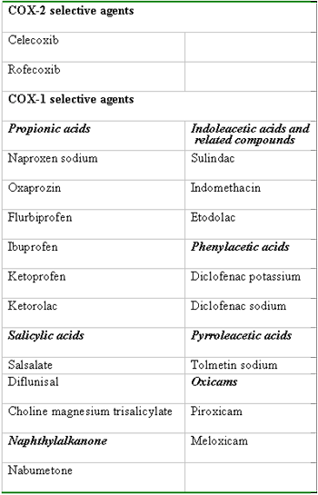
Patients who fail to respond should be considered candidates for nonsteroidal anti-inflammatory drug (NSAID) therapy. Different NSAIDs are about equally effective in relieving pain, and the choice of NSAID is usually influenced by side effect profile, dosing schedule and price. With the availability of new cyclooxygenase-2 (COX-2) inhibitors, which have an improved gastrointestinal safety profile, rheumatologists are recommending the older NSAIDs (COX-1 specific) less often. Recent evidence shows that COX-2 specific inhibitors have the same renal side effect profile as COX-1 specific NSAIDs and care must be exercised when these drugs are given to elderly patients who may have impaired kidney function.
Intraarticular steroid injections are used commonly in the management of OA, especially in the knee. Some evidence supports their benefit for one to three weeks but in practice many patients seem to have sustained benefit for up to six months.(14) Repeated injections more than three to four times a year have been discouraged because of concern about progressive cartilage damage.
Topical creams (e.g., 0.025% capsaicin cream) can be used in conjunction with the other treatment modalities but success is usually limited. Local application of cold packs, 2-3 times a day for 10-15 minutes can be used to supplement any treament regimen and helps decrease the inflammation in any given joint.(15)
Intraarticular hyaluronic acid has also been shown to be effective in some patients with knee OA. A recent review, however, concluded that usefulness of viscosupplementation by intraarticular injection of hyaluronic acid in the treatment of OA is promoted by the manufacturers and that few data exist to support this treatment. Further more, although some trials indicate that this modality results in relief of pain, similar results are also seen with placebo and it is not clear, even if statistically significant, that these results are clinically significant.(16)
Glucosamine sulfate has been available in different preparations for some time now and has been used by many patients for the treatment of their OA, especially of the knee. Until recently, there was a lack of evidence to support any role for glucosamine sulfate in the treatment of OA but newly published studies hint at possible benefit(17) but until further studies support these early findings, it is difficult to recommend this agent in the routine treatment of OA.
Magnets are commonly used by patients to help with the pain of OA. Even though they are probably safe, their efficacy for painful conditions like OA remains dubious because of limited research. Although there is a foundation of hope because of personal, anecdotal reports of success, much of the enthusiasm has been based on uncontrolled studies.(18) Larger studies should be conducted before magnets can be recommended to patients.
Surgical Treatment
Patients whose symptoms are not controlled with medical therapy and who have moderate to severe functional impairment are candidates for surgical management. Internal derangement of the knee can be treated with arthroscopic debridement and, if needed, meniscectomy. For young patients with knee OA, high tibial osteotomy could be used.
Total joint arthroplasty is a very effective treatment option for OA patients with severe disease nonresponsive to medical treatment, producing a dramatic improvement in their quality of life. Perioperative mortality is below 1% and short-term complications, like thromboembolic disease or infection, occur in fewer than 5% of cases. Porous coated prostheses, which allow for bone growth, help with fixation and decrease rates for loosening, thus reducing the need for revisions.
Table 1. Signs and Symptoms of OA.
| Symptoms | Signs |
| Joint pain | Limitation of range of motion |
| Loss of function | Crepitus |
| Morning stiffness | Bony enlargement |
| Gel phenomenon | Tenderness on palpation |
| Instability | Malalignment |
| Joint effusion |
Table 2. Prevalence of Osteoarthritis*.

Table 3. Radiologic Evidence of Knee OA from the Framingham Study.

Table 4. Commonly Used NSAIDs.
