Course Authors
Susan C. Stewart, M.D.
Release Date: 06/19/2000
Upon completion of this Cyberounds®, you should be able to:
Define the different types of lymphedema
Describe the anatomy and physiology of the lymphatic system
List the anatomic and physiologic features of the lymphatic system involved in Complete Decongestive Physiotherapy (CDP).
Lymphedema is the accumulation of fluid in the interstitial space because of defective transport by the lymphatic system. Inadequate development of the lymphatic system produces so-called primary lymphedema, while interruption of the lymphatics by infection, injury, surgery or radiation is classified as secondary lymphedema. Lymphedema may also occur as a consequence of the system overload present in certain obese patients. Another type of lymphedema can follow chronic venous insufficiency.
Epidemiology
Worldwide, the most common cause of lymphedema is infestation and damage by the parasitic disease, filariasis. In the United States, the most common cause in the upper extremities is axillary node dissection and radiation for breast cancer treatment. In the lower extremities, the most common types are congenital (i.e., primary) or lymphedema secondary to chronic venous insufficiency. In all of these cases, a disproportionately large number of women are affected. Other cancers treated with node dissection and radiation, like uterine, cervical and prostate, can be complicated by leg lymphedema. Malignant melanoma treatment is another frequent cause of lymphedema.(1),(4)
Morbidity
Because lymphedema in the U.S. is so often seen as a consequence of cancer treatment, it is easy to think of lymphedema in terms of what it is not. Unless it is a manifestation of a recurrence, it is not cancer. It is usually not life threatening. And we physicians often fall into the trap of thinking that lymphedema is not serious, and we shouldn't have to do anything about it. But let's talk about what lymphedema IS.
Lymphedema is a progressive, permanent, irreversible condition. It can cause substantial physical limitations, especially in the legs. There are complications, in particular, infections -- cellulitis, lymphangitis and septicemia -- that can be fatal. A recent study found that lymphedema increased the odds for developing erysipelas in the lower extremity 70-fold.(5) Lymphorrhea, which is blistering of the skin and exudation of fluid, opens a portal for infection, in addition to being malodorous and a therapeutic management challenge. The more massive the deformity, the more difficult it is to fit clothes, even to go out in public. For cancer patients, lymphedema becomes a constant reminder of their disease long after the incisions have healed and the hair grows back, and is a source of continuing psychological distress.
Treatment
Lymphedema patients do not like to be told, "It's not serious. Live with it." But until very recently in the U.S., there were no good treatments for lymphedema. Microsurgery, which involves anastomosing lymph vessels to veins, works initially but, subsequently, the connections fibrose and occlude the lumens. Though microsurgery is still being attempted in a few centers in different parts of the world, it is a lot of work and does not provide a long-term solution. Other types of surgery have generally been abandoned.(3)
Pneumatic pumps usually transfer fluid from the limb to the more proximal areas of the body quadrant drained by the involved lymph drainage system. When the pump is removed, the tissue fluid flows back into the limb. With continued use, pumps compress and damage the skin. Most patients find that the pumps do not help and stop using them before damage occurs. Amazingly, these devices are still covered by insurance policies, even though most patients find they are ineffective and can be detrimental.(6)
CDP, Complete Decongestive Physiotherapy, consists of a type of massage therapy called manual lymph drainage, followed by compression bandaging. Therapeutic exercises and meticulous skin care are also part of this program. CDP was brought to the U.S. in 1989 by Dr. Robert Lerner, formerly Chief of Surgery, Brooklyn Jewish Hospital, New York, after he visited the Foeldi Clinic in Germany and observed that the technique actually worked to control lymphedema.(7),(8)
The Anatomic Basis of Complete Decongestive Physiotherapy
Another term applied to this technique is Complex Decongestive Physiotherapy, and you may see variations on the two terms. Two key elements of the treatment, manual lymph drainage and compression bandaging, take advantage of unique anatomical features of the lymphatic system: the deep and superficial drainage vessels and the watershed connections between the body quadrants.
The smallest lymph vessels, situated in capillary beds, have a blind end and very thin walls of overlapping cells. These cells can be pulled apart by microfilaments to permit water, protein, tissue cells and microorganisms to pass into the vessel lumen.
Figure 1. Lymph Capillary & Blood Capillary.
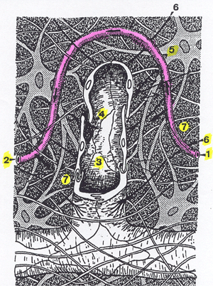
- Arterial side of the blood capillary
- Venous side of the blood capillary
- Lymph capillary
- Open junction "swinging flap"
- Fibrocyte
- Anchoring filaments
- Interstitial space
The content of the vessel is then called lymph. Valves in the vessels help retain and transport the lymph centrally, and, in the limbs, against gravity. The smallest vessels join together into larger and larger vessels. There is a very extensive network of lymph vessels in the skin called the dermal lymphatics, which is connected to the system that drains the muscles and other deeper tissues, called the deep lymphatics. In a normal circulation, the superficial system drains toward the deep system, which coalesces into vessels that are strained through the lymph nodes, then into the larger vessels that empty into major veins. A lymph node dissection will interrupt those larger vessels and if a critical number are destroyed, lymphedema will develop in the limb.
Figure 2. Main Lymphatic Trunks Entering Venous System.
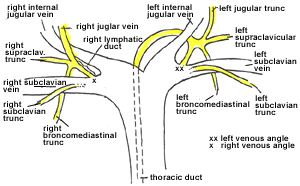
The thoracic duct transports lower extremity lymph to the left subclavian vein.
The other key anatomic fact is that the dermal lymphatic systems of the major body quadrants are connected by watersheds and anastomoses.
Figure 3. Lymphatic Watersheds and Anastomoses.
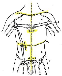
Major quadrant anastomoses are in yellow.
Manual Lymph Drainage
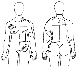
CDP takes advantage of the extensive connections between the superficial and deep lymphatic systems and the watershed areas in the following ways. The technique of manual lymph drainage over the trunk opens the watershed connections between the normal quadrants and the diseased quadrant. This type of massage is very gentle because it is directed toward the skin, not the muscles. The therapist must have a detailed knowledge of the direction of lymphatic flow in order to open anastomotic channels. The technique is then applied over the affected limb, working in a proximal to distal direction. Tissue fluid flows through the superficial lymphatics under the skin, bypassing the damaged node bearing areas and passing through the anastomoses to the normal node areas. For example, in the treatment of a lymphedematous arm, lymph would be directed to the contralateral axilla and the ipsilateral groin.
Figure 4. Manual Lymph Drainage Pathways for Left Arm Lymphedema.
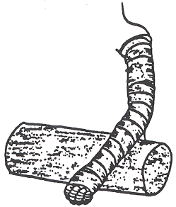
Bandaging and Compression Garments
The progress and the redirection of flow achieved by manual lymph drainage are maintained by bandaging the limb with multilayered bandages. During Phase I, or the intensive treatment program of CDP, a patient would receive manual lymph drainage and bandaging daily, once or twice a day, five days a week and remain bandaged between sessions.
Figure 5. Left Arm After Complete Bandaging.
Figures 1-5: With permission, Lerner Lymphedema Services Academy.
This treatment could continue as long as one or two weeks for a very mild case, or for up to 12 weeks for a very severe case. Part of the intensive treatment program is teaching the patient how to bandage independently for the maintenance program. Most limbs can be decompressed by at least 60% during the intensive program. The patient is then fitted with a compression garment for day wear. At night, the garment is removed and the limb bandaged. The home maintenance program is called Phase II.(2)
Figure 6. Mild Lymphedema, Left Arm.
Before 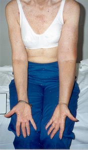 |
After 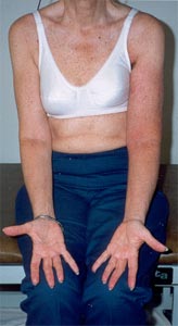 |
Note shiny, tense appearance of left forearm and thickened upper arm in the before treatment picture. The red appearance of the arm in the "after" picture is due to recent removal of bandages.
Figure 7. Severe Lymphedema, Right Arm.
Before
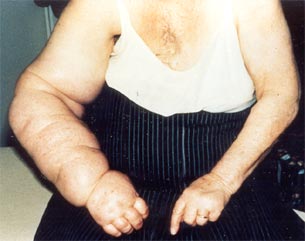
After
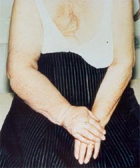
Before and after four weeks of treatment.
Figure 8. Left Leg Lymphedema in a Young Woman.
Before 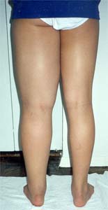 |
After 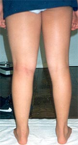 |
Before and after four weeks of treatment.
Figure 9. Severe Lymphedema of the Right Leg.
Before 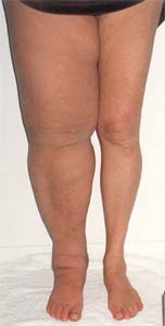 |
After 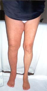 |
Before and after four weeks of treatment.
Figure 10. Varicose Veins, Chronic Venous Insufficiency and Lymphedema.
Before 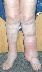 |
After 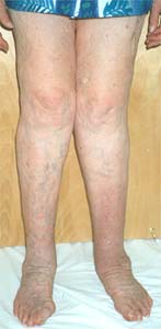 |
Stockings
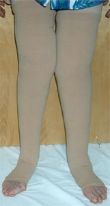
Before and after three weeks of treatment. This type of lymphedema responds very well to therapy. Removing the edema allows for application of appropriately sized support stockings. The picture on the right shows the patient in well-fitting, thigh-high stockings.
Figures 6-10: With permission, Lerner Lymphedema Services.
Therapeutic Exercises
This part of the program takes advantage of another anatomic feature of the lymphatic system -- the dense network of lymphatic vessels on the muscle fascia. This network is connected to the dermal lymphatics. When the muscles contract, lymphatic vessels contract as well and the lymph flows faster. The exercises must be performed with the bandages in place because compression is required to maintain dermal lymph flow. If physical exercise is undertaken without compression, the increased blood flow will produce more interstitial fluid and the edema will increase because the dermal lymphatics require counter pressure (otherwise, the skin and subcutaneous tissue will just expand to accommodate the fluid). The best form of exercise for lymphedema patients is swimming, or other water exercise, because the water provides tissue counter pressure and the muscular activity causes the lymph to flow.
Skin Care
The most serious complication of lymphedema is infection. Minor injuries can precipitate major episodes of cellulitis, lymphangitis or septicemia. Inflammation of the lymph vessels, lymphangitis, causes dilatation and incompetence of the vessels. Inflammation and infection can destroy the valves and the vessels themselves, which will further intensify the case of lymphedema. Indeed, some patients present for the first time with lymphedema after an episode of cellulitis.
In CDP, the skin of the affected limb is treated with a low pH lotion that inhibits bacterial growth. Arm patients are cautioned to use gloves for gardening, dishwashing or any activity that would expose them to cuts. "Professional" manicures and removal of the nail cuticles should not be done. Leg patients should not walk in bare feet. For outdoor activities, the skin should be protected with liberal sunscreen. Patients are instructed to look for the first signs of cellulitis and immediately start an antibiotic, which should be kept on hand at all times.
Timing of CDP
CDP works best when it is started early in the development of lymphedema. I don't advise waiting until the condition is noticeable and causing problems -- an early consultation is very useful so that patients can evaluate their options.
CDP is not for everyone. In fact, it takes a very determined, dedicated patient to use this treatment to its greatest advantage. My experience has been that when a patient experiences a noticeable change in her lymphedema problem, or sees that what appeared to be an uncontrollable condition can be controlled, she becomes motivated to continue the treatment on the maintenance program.
Summary
Lymphedema is a chronic, incurable condition resulting from inadequate lymph drainage, almost always of an arm or a leg. It can be congenital or acquired, most frequently resulting from cancer surgery. The safest and most effective treatment is a technique, relatively new to the United States, called Complete (or Complex) Decongestive Therapy (or Physiotherapy). Patients should be referred for this type of treatment as early as possible in the course of developing lymphedema.
With appreciation to Dr. Robert Lerner for reviewing this article and for permission to use his figures and photos.
Websites
Lerner Lymphedema Services - lymphedema treatment centers and therapist training academy.
National Lymphedema Network - support groups, newsletter, information about treatment centers and training programs.